 Explore
 Validate
 Learn
Learn Western blot
Antibody data
- Antibody Data
- Antigen structure
- References [10]
- Comments [0]
- Validations
- Western blot [2]
- Immunocytochemistry [1]
- Immunohistochemistry [2]
- Other assay [1]
Submit
Validation data
Reference
Comment
Report error
- Product number
- MA5-12547 - Provider product page

- Provider
- Invitrogen Antibodies
- Product name
- E-cadherin Monoclonal Antibody (NCH-38)
- Antibody type
- Monoclonal
- Antigen
- Recombinant full-length protein
- Description
- MA5-12547 targets E-Cadherin in IHC (P) applications and shows reactivity with Human samples. The MA5-12547 immunogen is e-cadherin and GST recombinant protein.
- Reactivity
- Human, Mouse
- Host
- Mouse
- Isotype
- IgG
- Antibody clone number
- NCH-38
- Vial size
- 500 µL
- Concentration
- Conc. Not Determined
- Storage
- 4° C
Submitted references Overexpression and Tyr421-phosphorylation of cortactin is induced by three-dimensional spheroid culturing and contributes to migration and invasion of pancreatic ductal adenocarcinoma (PDAC) cells.
Ankyrin-G regulated epithelial phenotype is required for mouse lens morphogenesis and growth.
Acetyl-CoA carboxylase inhibitors attenuate WNT and Hedgehog signaling and suppress pancreatic tumor growth.
Malignant transformation of adenomyoepithelioma of the breast by a monophasic population: a report of two cases and review of literature.
Five primary human pancreatic adenocarcinoma cell lines established by the outgrowth method.
The expression of cytoskeleton regulatory protein Mena in colorectal lesions.
Nogo-A inhibitory peptide (NEP1-40) increases pan-cadherin expression following mild cortical contusion injury in rats.
The expression of cytokeratin 5/6 in invasive lobular carcinoma of the breast: evidence of a basal-like subset?
Human ovarian surface epithelial cells immortalized with hTERT maintain functional pRb and p53 expression.
Cystic hypersecretory carcinoma: rare and poorly recognized variant of intraductal carcinoma of the breast. Report of five cases.
Stock K, Borrink R, Mikesch JH, Hansmeier A, Rehkämper J, Trautmann M, Wardelmann E, Hartmann W, Sperveslage J, Steinestel K
Cancer cell international 2019;19:77
Cancer cell international 2019;19:77
Ankyrin-G regulated epithelial phenotype is required for mouse lens morphogenesis and growth.
Rasiah PK, Maddala R, Bennett V, Rao PV
Developmental biology 2019 Feb 1;446(1):119-131
Developmental biology 2019 Feb 1;446(1):119-131
Acetyl-CoA carboxylase inhibitors attenuate WNT and Hedgehog signaling and suppress pancreatic tumor growth.
Petrova E, Scholz A, Paul J, Sturz A, Haike K, Siegel F, Mumberg D, Liu N
Oncotarget 2017 Jul 25;8(30):48660-48670
Oncotarget 2017 Jul 25;8(30):48660-48670
Malignant transformation of adenomyoepithelioma of the breast by a monophasic population: a report of two cases and review of literature.
Marian C, Boila A, Soanca D, Malau M, Podeanu DM, Resetkova E, Stolnicu S
APMIS : acta pathologica, microbiologica, et immunologica Scandinavica 2013 Apr;121(4):272-9
APMIS : acta pathologica, microbiologica, et immunologica Scandinavica 2013 Apr;121(4):272-9
Five primary human pancreatic adenocarcinoma cell lines established by the outgrowth method.
Rückert F, Aust D, Böhme I, Werner K, Brandt A, Diamandis EP, Krautz C, Hering S, Saeger HD, Grützmann R, Pilarsky C
The Journal of surgical research 2012 Jan;172(1):29-39
The Journal of surgical research 2012 Jan;172(1):29-39
The expression of cytoskeleton regulatory protein Mena in colorectal lesions.
Gurzu S, Jung I, Prantner I, Ember I, Pávai Z, Mezei T
Romanian journal of morphology and embryology = Revue roumaine de morphologie et embryologie 2008;49(3):345-9
Romanian journal of morphology and embryology = Revue roumaine de morphologie et embryologie 2008;49(3):345-9
Nogo-A inhibitory peptide (NEP1-40) increases pan-cadherin expression following mild cortical contusion injury in rats.
Atalay B, Bavbek M, Ozen O, Nacar A, Gülşen S, Yiğitkanli K, Caner H, Altinörs N
Turkish neurosurgery 2008 Oct;18(4):356-65
Turkish neurosurgery 2008 Oct;18(4):356-65
The expression of cytokeratin 5/6 in invasive lobular carcinoma of the breast: evidence of a basal-like subset?
Fadare O, Wang SA, Hileeto D
Human pathology 2008 Mar;39(3):331-6
Human pathology 2008 Mar;39(3):331-6
Human ovarian surface epithelial cells immortalized with hTERT maintain functional pRb and p53 expression.
Li NF, Broad S, Lu YJ, Yang JS, Watson R, Hagemann T, Wilbanks G, Jacobs I, Balkwill F, Dafou D, Gayther SA
Cell proliferation 2007 Oct;40(5):780-94
Cell proliferation 2007 Oct;40(5):780-94
Cystic hypersecretory carcinoma: rare and poorly recognized variant of intraductal carcinoma of the breast. Report of five cases.
Skalova A, Ryska A, Kajo K, Di Palma S, Kinkor Z, Michal M
Histopathology 2005 Jan;46(1):43-9
Histopathology 2005 Jan;46(1):43-9
No comments: Submit comment
Supportive validation
- Submitted by
- Invitrogen Antibodies (provider)
- Main image

- Experimental details
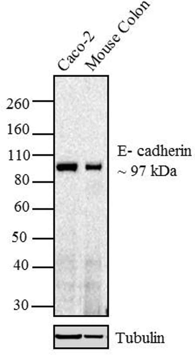
- Western blot analysis of E-Cadherin was performed using whole cell extract and tissue lysate of Caco-2 (Lane 1) and Mouse Colon (Lane 2). The blot was probed with Anti-E-cadherin Mouse Monoclonal Antibody (Product # MA5-12547, 1:250 dilution) and detected by chemiluminescence using Goat anti-Mouse IgG (H+L) Superclonal™ Secondary Antibody, HRP conjugate (Product # A28177, 0.4 µg/mL, 1:2500 dilution). A 97 kDa band corresponding to E-Cadherin was observed across cell lines tested. Known quantity of protein samples were electrophoresed using Novex® NuPAGE® 4-12 % Bis-Tris gel (Product # NP0321BOX), XCell SureLock™ Electrophoresis System (Product # EI0002) and Novex® Sharp Pre-Stained Protein Standard (Product # LC5800). Resolved proteins were then transferred onto a nitrocellulose membrane by overnight transfer method. The membrane was probed with the relevant primary and secondary Antibody using iBind™ Flex Western Starter Kit (Product # SLF2000S). Chemiluminescent detection was performed using Pierce™ ECL Western Blotting Substrate (Product # 32106).
- Submitted by
- Invitrogen Antibodies (provider)
- Main image

- Experimental details
- Western blot was performed using Anti-E-cadherin Monoclonal Antibody (NCH-38)(Product # MA5-12547) and a 110kDa band corresponding to E-cadherin was observed across in all tested cell lines and tissues, except MDA-MB-231, SW480 and BJ. Whole cell extracts (30 µg lysate) of MCF7 (Lane 1), MDA-MB-231 (Lane 2), SW480 (Lane 3), Caco-2 (Lane 4), BJ (Lane 5), Mouse Stomach (Lane 6), Rat Stomach (Lane 7) were electrophoresed using NuPAGE™ 4-12% Bis-Tris Protein Gel (Product # NP0322BOX). Resolved proteins were then transferred onto a Nitrocellulose membrane (Product # IB23001) by iBlot® 2 Dry Blotting System (Product # IB21001). The blot was probed with the primary antibody (1:250) and detected by chemiluminescence with Goat anti-Mouse IgG (H+L) Superclonal™ Recombinant Secondary Antibody, HRP (Product # A28177,1:4000) using the iBright FL 1000 (Product # A32752). Chemiluminescent detection was performed using SuperSignal™ West Dura Extended Duration Substrate (Product # 34076). The additional bands at ~80kDa represent proteolytic cleavage products of E-cadherin.

Supportive validation
- Submitted by
- Invitrogen Antibodies (provider)
- Main image

- Experimental details
- Immunofluorescence analysis of E-cadherin was performed using 70 percent confluent log phase Caco-2 cells. The cells were fixed with 4% paraformaldehyde for 10 minutes, permeabilized with 0.1% Triton™ X-100 for 15 minutes, and blocked with 2% BSA for 45 minutes at room temperature. The cells were labeled with E-cadherin Monoclonal Antibody (NCH-38) (Product # MA5-12547) at 1:200 in 0.1% BSA, incubated at 4 degree celsius overnight and then labeled with Goat anti-Mouse IgG (H+L) Highly Cross-Adsorbed Secondary Antibody, Alexa Fluor Plus 488 (Product # A32723), (1:2000), for 45 minutes at room temperature (Panel a: Green). Nuclei (Panel b: Blue) were stained with ProLong™ Diamond Antifade Mountant with DAPI (Product # P36962). F-actin (Panel c: Red) was stained with Rhodamine Phalloidin (Product # R415, 1:300). Panel d represents the merged image showing plasma membrane localization. Panel e represents BJ cells, showing no expression of E-cadherin. Panel f represents control cells with no primary antibody to assess background. The images were captured at 60X magnification.

Supportive validation
- Submitted by
- Invitrogen Antibodies (provider)
- Main image

- Experimental details
- Formalin-fixed, paraffin-embedded human breast carcinoma stained with E-Cadherin antibody using peroxidase-conjugate and AEC chromogen. Note membrane staining of tumor cells.

- Submitted by
- Invitrogen Antibodies (provider)
- Main image

- Experimental details
- Immunohistochemistry analysis of E-cadherin/CDH1 (NCH-38) showing staining in the membrane and weak cytoplasm of paraffin-embedded human breast carcinoma (right) compared to a negative control without primary antibody (left). To expose target proteins, antigen retrieval was performed using 10mM sodium citrate (pH 6.0), microwaved for 8-15 min. Following antigen retrieval, tissues were blocked in 3% H2O2-methanol for 15 min at room temperature, washed with ddH2O and PBS, and then probed with a E-cadherin/CDH1 Antibody (NCH-38) Mouse Monoclonal Antibody (Product # MA5-12547) diluted in 3% BSA-PBS at a dilution of 1:20 for 1 hour at 37°C in a humidified chamber. Tissues were washed extensively in PBST and detection was performed using an HRP-conjugated secondary antibody followed by colorimetric detection using a DAB kit. Tissues were counterstained with hematoxylin and dehydrated with ethanol and xylene to prep for mounting.

Supportive validation
- Submitted by
- Invitrogen Antibodies (provider)
- Main image

- Experimental details
- Figure 3 BAY ACC002 Decreases WNT and HH Signaling in Pancreatic Cancer Models In Vivo A . AXIN2 and GLI1 expression in Capan-2 pancreatic cancer tumors, treated with vehicle or 30 mg/kg/day BAY ACC002 for 7 days. RNA was extracted from the tumors and expression of AXIN2 and GLI1 was determined by qRT-PCR. Each bar represents mean+-SEM, with individual animals represented by dots ( n = 4, *, p < 0.05). B . IHC analysis of beta-catenin expression in representative pancreatic cancer sections from mice, carrying Capan-2 or PAXF 2046 tumors, treated with vehicle or BAY ACC002 (35 mg/kg/day on days 1-5, and then 30 mg/kg/day on a 3 days ON/1 day OFF schedule until day 35 for the Capan-2 model; and 30 mg/kg/day for 29 days for the PAXF 2046 model). Arrows point to nuclear beta-catenin expression. C . IHC analysis of GLI1 expression in representative pancreatic cancer sections from Capan-2 and PAXF 2046 tumors described in (B). Arrows point to GLI1 positive cells. D . H&E, Alcian blue, and E-cadherin IHC staining of representative sections from the PAXF 2046 tumors, described in (B). Scale bar for (B), (C), and the third set of (D) is 50 mum, and for the first two sets of (D), 100 mum.
