Explore
 Validate
 Learn
Learn Western blot
Antibody data
- Antibody Data
- Antigen structure
- References [20]
- Comments [0]
- Validations
- Western blot [2]
- Immunocytochemistry [2]
- Immunohistochemistry [1]
- Flow cytometry [1]
Submit
Validation data
Reference
Comment
Report error
- Product number
- AF648 - Provider product page

- Provider
- R&D Systems
- Product name
- Human/Mouse E-Cadherin Antibody
- Antibody type
- Polyclonal
- Description
- Antigen Affinity-purified. Detects human E-Cadherin in direct ELISAs and Western blots.
- Reactivity
- Human, Mouse
- Host
- Goat
- Conjugate
- Unconjugated
- Antigen sequence
P12830- Isotype
- IgG
- Vial size
- 100 ug
- Concentration
- LYOPH
- Storage
- Use a manual defrost freezer and avoid repeated freeze-thaw cycles. 12 months from date of receipt, -20 to -70 °C as supplied. 1 month, 2 to 8 °C under sterile conditions after reconstitution. 6 months, -20 to -70 °C under sterile conditions after reconstitution.
Submitted references Enhanced Utilization of Induced Pluripotent Stem Cell-Derived Human Intestinal Organoids Using Microengineered Chips.
Identification of Piperazinylbenzenesulfonamides as New Inhibitors of Claudin-1 Trafficking and Hepatitis C Virus Entry.
A novel transforming growth factor beta-induced long noncoding RNA promotes an inflammatory microenvironment in human intrahepatic cholangiocarcinoma.
Placenta-specific protein 1 promotes cell proliferation and invasion in non-small cell lung cancer.
Acoustic Tweezing Cytometry Induces Rapid Initiation of Human Embryonic Stem Cell Differentiation.
Directed Differentiation of Human Induced Pluripotent Stem Cells into Fallopian Tube Epithelium.
A process engineering approach to increase organoid yield.
Novel role for IL-22 in protection during chronic Mycobacterium tuberculosis HN878 infection.
miR-151a induces partial EMT by regulating E-cadherin in NSCLC cells.
Wnt/β-catenin promotes gastric fundus specification in mice and humans.
Surface Topography Guides Morphology and Spatial Patterning of Induced Pluripotent Stem Cell Colonies.
Canine spontaneous head and neck squamous cell carcinomas represent their human counterparts at the molecular level.
Molecular homology and difference between spontaneous canine mammary cancer and human breast cancer.
Novel human renal proximal tubular cell line for the production of complex proteins.
Penton-dodecahedral particles trigger opening of intercellular junctions and facilitate viral spread during adenovirus serotype 3 infection of epithelial cells.
Interleukin-15 plays a central role in human kidney physiology and cancer through the γc signaling pathway.
Snail involves in the transforming growth factor β1-mediated epithelial-mesenchymal transition of retinal pigment epithelial cells.
Tissue engineering of oral dysplasia.
Downregulation of Rap1GAP contributes to Ras transformation.
Requirement for intercellular adhesion molecule 1 and caveolae in invasion of human oral epithelial cells by Porphyromonas gingivalis.
Workman MJ, Gleeson JP, Troisi EJ, Estrada HQ, Kerns SJ, Hinojosa CD, Hamilton GA, Targan SR, Svendsen CN, Barrett RJ
Cellular and molecular gastroenterology and hepatology 2018;5(4):669-677.e2
Cellular and molecular gastroenterology and hepatology 2018;5(4):669-677.e2
Identification of Piperazinylbenzenesulfonamides as New Inhibitors of Claudin-1 Trafficking and Hepatitis C Virus Entry.
Riva L, Song OR, Prentoe J, Helle F, L'homme L, Gattolliat CH, Vandeputte A, Fénéant L, Belouzard S, Baumert TF, Asselah T, Bukh J, Brodin P, Cocquerel L, Rouillé Y, Dubuisson J
Journal of virology 2018 May 15;92(10)
Journal of virology 2018 May 15;92(10)
A novel transforming growth factor beta-induced long noncoding RNA promotes an inflammatory microenvironment in human intrahepatic cholangiocarcinoma.
Merdrignac A, Angenard G, Allain C, Petitjean K, Bergeat D, Bellaud P, Fautrel A, Turlin B, Clément B, Dooley S, Sulpice L, Boudjema K, Coulouarn C
Hepatology communications 2018 Mar;2(3):254-269
Hepatology communications 2018 Mar;2(3):254-269
Placenta-specific protein 1 promotes cell proliferation and invasion in non-small cell lung cancer.
Yang L, Zha TQ, He X, Chen L, Zhu Q, Wu WB, Nie FQ, Wang Q, Zang CS, Zhang ML, He J, Li W, Jiang W, Lu KH
Oncology reports 2018 Jan;39(1):53-60
Oncology reports 2018 Jan;39(1):53-60
Acoustic Tweezing Cytometry Induces Rapid Initiation of Human Embryonic Stem Cell Differentiation.
Topal T, Hong X, Xue X, Fan Z, Kanetkar N, Nguyen JT, Fu J, Deng CX, Krebsbach PH
Scientific reports 2018 Aug 28;8(1):12977
Scientific reports 2018 Aug 28;8(1):12977
Directed Differentiation of Human Induced Pluripotent Stem Cells into Fallopian Tube Epithelium.
Yucer N, Holzapfel M, Jenkins Vogel T, Lenaeus L, Ornelas L, Laury A, Sareen D, Barrett R, Karlan BY, Svendsen CN
Scientific reports 2017 Sep 6;7(1):10741
Scientific reports 2017 Sep 6;7(1):10741
A process engineering approach to increase organoid yield.
Arora N, Imran Alsous J, Guggenheim JW, Mak M, Munera J, Wells JM, Kamm RD, Asada HH, Shvartsman SY, Griffith LG
Development (Cambridge, England) 2017 Mar 15;144(6):1128-1136
Development (Cambridge, England) 2017 Mar 15;144(6):1128-1136
Novel role for IL-22 in protection during chronic Mycobacterium tuberculosis HN878 infection.
Treerat P, Prince O, Cruz-Lagunas A, Muñoz-Torrico M, Salazar-Lezama MA, Selman M, Fallert-Junecko B, Reinhardt TA, Alcorn JF, Kaushal D, Zuñiga J, Rangel-Moreno J, Kolls JK, Khader SA
Mucosal immunology 2017 Jul;10(4):1069-1081
Mucosal immunology 2017 Jul;10(4):1069-1081
miR-151a induces partial EMT by regulating E-cadherin in NSCLC cells.
Daugaard I, Sanders KJ, Idica A, Vittayarukskul K, Hamdorf M, Krog JD, Chow R, Jury D, Hansen LL, Hager H, Lamy P, Choi CL, Agalliu D, Zisoulis DG, Pedersen IM
Oncogenesis 2017 Jul 31;6(7):e366
Oncogenesis 2017 Jul 31;6(7):e366
Wnt/β-catenin promotes gastric fundus specification in mice and humans.
McCracken KW, Aihara E, Martin B, Crawford CM, Broda T, Treguier J, Zhang X, Shannon JM, Montrose MH, Wells JM
Nature 2017 Jan 12;541(7636):182-187
Nature 2017 Jan 12;541(7636):182-187
Surface Topography Guides Morphology and Spatial Patterning of Induced Pluripotent Stem Cell Colonies.
Abagnale G, Sechi A, Steger M, Zhou Q, Kuo CC, Aydin G, Schalla C, Müller-Newen G, Zenke M, Costa IG, van Rijn P, Gillner A, Wagner W
Stem cell reports 2017 Aug 8;9(2):654-666
Stem cell reports 2017 Aug 8;9(2):654-666
Canine spontaneous head and neck squamous cell carcinomas represent their human counterparts at the molecular level.
Liu D, Xiong H, Ellis AE, Northrup NC, Dobbin KK, Shin DM, Zhao S
PLoS genetics 2015 Jun;11(6):e1005277
PLoS genetics 2015 Jun;11(6):e1005277
Molecular homology and difference between spontaneous canine mammary cancer and human breast cancer.
Liu D, Xiong H, Ellis AE, Northrup NC, Rodriguez CO Jr, O'Regan RM, Dalton S, Zhao S
Cancer research 2014 Sep 15;74(18):5045-56
Cancer research 2014 Sep 15;74(18):5045-56
Novel human renal proximal tubular cell line for the production of complex proteins.
Fliedl L, Manhart G, Kast F, Katinger H, Kunert R, Grillari J, Wieser M, Grillari-Voglauer R
Journal of biotechnology 2014 Apr 20;176:29-39
Journal of biotechnology 2014 Apr 20;176:29-39
Penton-dodecahedral particles trigger opening of intercellular junctions and facilitate viral spread during adenovirus serotype 3 infection of epithelial cells.
Lu ZZ, Wang H, Zhang Y, Cao H, Li Z, Fender P, Lieber A
PLoS pathogens 2013 Oct;9(10):e1003718
PLoS pathogens 2013 Oct;9(10):e1003718
Interleukin-15 plays a central role in human kidney physiology and cancer through the γc signaling pathway.
Giron-Michel J, Azzi S, Khawam K, Mortier E, Caignard A, Devocelle A, Ferrini S, Croce M, François H, Lecru L, Charpentier B, Chouaib S, Azzarone B, Eid P
PloS one 2012;7(2):e31624
PloS one 2012;7(2):e31624
Snail involves in the transforming growth factor β1-mediated epithelial-mesenchymal transition of retinal pigment epithelial cells.
Li H, Wang H, Wang F, Gu Q, Xu X
PloS one 2011;6(8):e23322
PloS one 2011;6(8):e23322
Tissue engineering of oral dysplasia.
Gaballah K, Costea DE, Hills A, Gollin SM, Harrison P, Partridge M
The Journal of pathology 2008 Jul;215(3):280-9
The Journal of pathology 2008 Jul;215(3):280-9
Downregulation of Rap1GAP contributes to Ras transformation.
Tsygankova OM, Prendergast GV, Puttaswamy K, Wang Y, Feldman MD, Wang H, Brose MS, Meinkoth JL
Molecular and cellular biology 2007 Oct;27(19):6647-58
Molecular and cellular biology 2007 Oct;27(19):6647-58
Requirement for intercellular adhesion molecule 1 and caveolae in invasion of human oral epithelial cells by Porphyromonas gingivalis.
Tamai R, Asai Y, Ogawa T
Infection and immunity 2005 Oct;73(10):6290-8
Infection and immunity 2005 Oct;73(10):6290-8
No comments: Submit comment
Supportive validation
- Submitted by
- R&D Systems (provider)
- Main image
- Experimental details
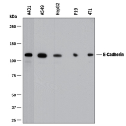
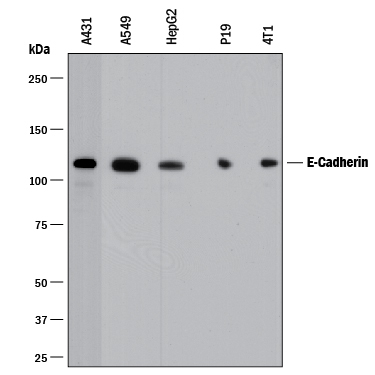
- Detection of Human and Mouse E-Cadherin by Western Blot. Western blot shows lysates of A431 human epithelial carcinoma cell line, A549 human lung carcinoma cell line, HepG2 human hepatocellular carcinoma cell line, P19 mouse embryonal carcinoma cell line, and 4T1 mouse breast cancer cell line. PVDF membrane was probed with 0.5 µg/mL of Goat Anti-Human/Mouse E-Cadherin Antigen Affinity-purified Polyclonal Antibody (Catalog # AF648) followed by HRP-conjugated Anti-Goat IgG Secondary Antibody (Catalog # HAF017). A specific band was detected for E-Cadherin at approximately 110 kDa (as indicated). This experiment was conducted under reducing conditions and using Immunoblot Buffer Group 1.
- Submitted by
- R&D Systems (provider)
- Main image

- Experimental details
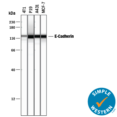
- Detection of Human and Mouse E-Cadherin by Simple WesternTM. Simple Western lane view shows lysates of 4T1 mouse breast cancer cell line, P19 mouse embryonal carcinoma cell line, A431 human epithelial carcinoma cell line, and MCF-7 human breast cancer cell line, loaded at 0.2 mg/mL. A specific band was detected for E-Cadherin at approximately 128 kDa (as indicated) using 5 µg/mL of Goat Anti-Human/Mouse E-Cadherin Antigen Affinity-purified Polyclonal Antibody (Catalog # AF648) followed by 1:50 dilution of HRP-conjugated Anti-Goat IgG Secondary Antibody (Catalog # HAF017). This experiment was conducted under reducing conditions and using the 12-230 kDa separation system.
Supportive validation
- Submitted by
- R&D Systems (provider)
- Main image

- Experimental details
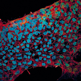
- E-Cadherin and SOX2 in BG01V Human Stem Cells. E-Cadherin and SOX2 were detected in BG01V human embryonic stem cells using 10 µg/mL Goat Anti-Human/Mouse E-Cadherin Antigen Affinity-purified Polyclonal Antibody (Catalog # AF648) and 10 µg/mL Human/Mouse SOX2 Monoclonal Antibody (Catalog # MAB2018). Cells were incubated with primary antibodies for 3 hours at room temperature. Cells were stained for E-Cadherin using the NorthernLights™ 557-conjugated Anti-Goat IgG Secondary Antibody (green; Catalog # NL001) and for SOX2 using the NorthernLights 493-conjugated Anti-Mouse Secondary Antibody (red; Catalog # NL009). Cells were counterstained with DAPI (blue). View our protocol for Fluorescent ICC Staining of Cells on Coverslips.
- Submitted by
- R&D Systems (provider)
- Main image

- Experimental details
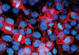
- E-Cadherin in Human Epidermoid Carcinoma Cells. E-Cadherin was detected in immersion fixed human epidermoid carcinoma cells using 10 µg/mL Goat Anti-Human/Mouse E-Cadherin Antigen Affinity-purified Polyclonal Antibody (Catalog # AF648) for 3 hours at room temperature. Cells were stained with the NorthernLights™ 557-conjugated Anti-Goat IgG Secondary Antibody (red; Catalog # NL001) and counterstained with DAPI (blue). View our protocol for Fluorescent ICC Staining of Cells on Coverslips.
Supportive validation
- Submitted by
- R&D Systems (provider)
- Main image
- Experimental details
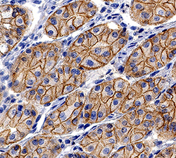
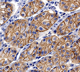
- E-Cadherin in Human Stomach. E-Cadherin was detected in immersion fixed paraffin-embedded sections of human stomach using Goat Anti-Human/Mouse E-Cadherin Antigen Affinity-purified Polyclonal Antibody (Catalog # AF648) at 0.3 µg/mL for 1 hour at room temperature followed by incubation with the Anti-Goat IgG VisUCyte™ HRP Polymer Antibody (Catalog # VC004). Tissue was stained using DAB (brown) and counterstained with hematoxylin (blue). Specific staining was localized to cell membrane and cytoplasm in gastric glands. View our protocol for IHC Staining with VisUCyte HRP Polymer Detection Reagents.
Supportive validation
- Submitted by
- R&D Systems (provider)
- Main image

- Experimental details
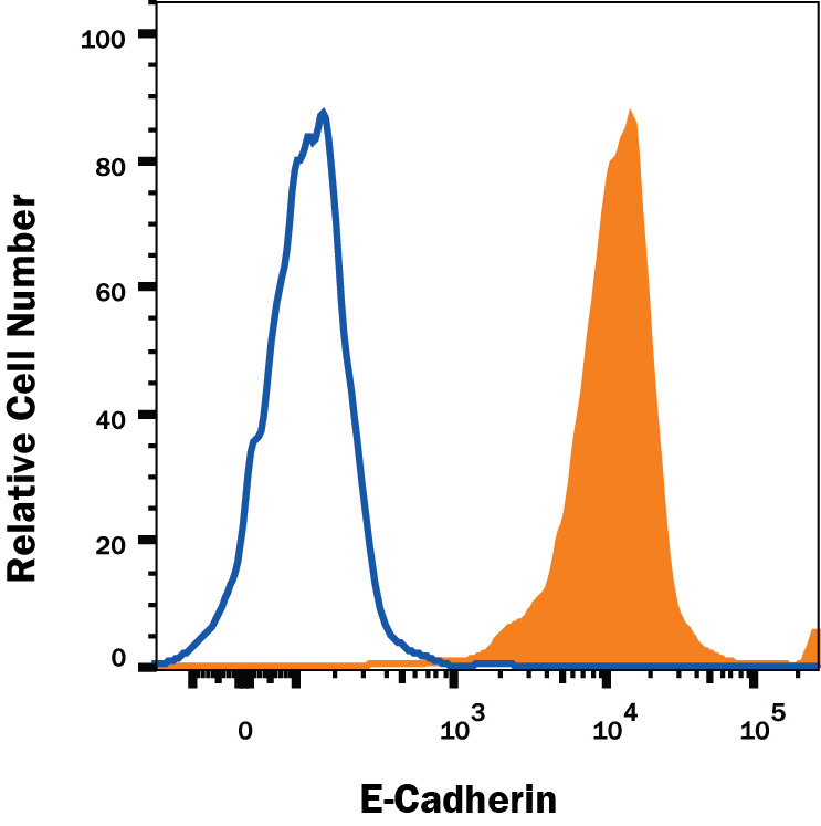
- Detection of E-Cadherin in MCF-7 Human Cell Line by Flow Cytometry. MCF-7 human breast cancer cell line was stained with Goat Anti-Human/Mouse E-Cadherin Antigen Affinity-purified Polyclonal Antibody (Catalog # AF648, filled histogram) or isotype control antibody (Catalog # AB-108-C, open histogram), followed by Allophycocyanin-conjugated Anti-Goat IgG Secondary Antibody (Catalog # F0108). View our protocol for Staining Membrane-associated Proteins.