 Explore
 Validate
 Learn
Learn Western blot
 Immunohistochemistry
Antibody data
- Antibody Data
- Antigen structure
- References [21]
- Comments [0]
- Validations
- Immunohistochemistry [2]
- Flow cytometry [5]
Submit
Validation data
Reference
Comment
Report error
- Product number
- NB110-68136 - Provider product page

- Provider
- Novus Biologicals
- Proper citation
- Novus Cat#NB110-68136, RRID:AB_1110904
- Product name
- Mouse Monoclonal Tenascin C Antibody
- Antibody type
- Monoclonal
- Description
- Protein A purified. NB110-68136 specifically reacts with Domain B on FNIII repeats of Tenascin C.
- Reactivity
- Human, Mouse, Rat, Feline
- Host
- Mouse
- Isotype
- IgG
- Vial size
- 0.1 ml
- Concentration
- 1.0 mg/ml
- Storage
- Store at 4C short term. Aliquot and store at -20C long term. Avoid freeze-thaw cycles.
Submitted references Increased expression of periostin and tenascin-C in eyes with neovascular glaucoma secondary to PDR.
Fibroblasts from the Human Skin Dermo-Hypodermal Junction are Distinct from Dermal Papillary and Reticular Fibroblasts and from Mesenchymal Stem Cells and Exhibit a Specific Molecular Profile Related to Extracellular Matrix Organization and Modeling.
Targeted disruption of the iNOS gene improves adipose tissue inflammation and fibrosis in leptin-deficient ob/ob mice: role of tenascin C.
Novel role of CCN3 that maintains the differentiated phenotype of articular cartilage.
Tenascin-C drives persistence of organ fibrosis.
Infrared LED light therapy influences the expression of fibronectin and tenascin in skin wounds of malnourished rats--a preliminary study.
Direct progression of capsular invasive carcinomas from subcapsular focal hyperplasias induced by hypothyroidism-mediated tumor promotion in a rat two-stage thyroid carcinogenesis model.
VARA attenuates hyperoxia-induced impaired alveolar development and lung function in newborn mice.
Promoting effects of carminic acid-enriched cochineal extracts on capsular invasive thyroid carcinomas through targeting activation of angiogenesis in rats.
Promoting effects of carminic acid-enriched cochineal extracts on capsular invasive thyroid carcinomas through targeting activation of angiogenesis in rats.
Lac color inhibits development of rat thyroid carcinomas through targeting activation of plasma hyaluronan-binding protein.
Novel effects of CCN3 that may direct the differentiation of chondrocytes.
Increased presence of stromal myofibroblasts and tenascin-C with malignant progression in canine mammary tumors.
A migration signature and plasma biomarker panel for pancreatic adenocarcinoma.
Regeneration of the perineurium after microsurgical resection examined with immunolabeling for tenascin-C and alpha smooth muscle actin.
Expression of tenascin-C and its isoforms in the breast.
B and C domain containing tenascin-C: urinary markers for invasiveness of urothelial carcinoma of the urinary bladder?
Expression of large tenascin-C splice variants by hepatic stellate cells/myofibroblasts in chronic hepatitis C.
Switch to an invasive growth phase in melanoma is associated with tenascin-C, fibronectin, and procollagen-I forming specific channel structures for invasion.
Tenascin-C regulates recruitment of myofibroblasts during tissue repair after myocardial injury.
Involvement of large tenascin-C splice variants in breast cancer progression.
Ishikawa K, Kohno RI, Mori K, Murakami Y, Nakao S, Akiyama M, Yoshida S, Sonoda KH
Graefe's archive for clinical and experimental ophthalmology = Albrecht von Graefes Archiv fur klinische und experimentelle Ophthalmologie 2020 Mar;258(3):621-628
Graefe's archive for clinical and experimental ophthalmology = Albrecht von Graefes Archiv fur klinische und experimentelle Ophthalmologie 2020 Mar;258(3):621-628
Fibroblasts from the Human Skin Dermo-Hypodermal Junction are Distinct from Dermal Papillary and Reticular Fibroblasts and from Mesenchymal Stem Cells and Exhibit a Specific Molecular Profile Related to Extracellular Matrix Organization and Modeling.
Haydont V, Neiveyans V, Perez P, Busson É, Lataillade J, Asselineau D, Fortunel NO
Cells 2020 Feb 5;9(2)
Cells 2020 Feb 5;9(2)
Targeted disruption of the iNOS gene improves adipose tissue inflammation and fibrosis in leptin-deficient ob/ob mice: role of tenascin C.
Becerril S, Rodríguez A, Catalán V, Méndez-Giménez L, Ramírez B, Sáinz N, Llorente M, Unamuno X, Gómez-Ambrosi J, Frühbeck G
International journal of obesity (2005) 2018 Aug;42(8):1458-1470
International journal of obesity (2005) 2018 Aug;42(8):1458-1470
Novel role of CCN3 that maintains the differentiated phenotype of articular cartilage.
Janune D, Abd El Kader T, Aoyama E, Nishida T, Tabata Y, Kubota S, Takigawa M
Journal of bone and mineral metabolism 2017 Nov;35(6):582-597
Journal of bone and mineral metabolism 2017 Nov;35(6):582-597
Tenascin-C drives persistence of organ fibrosis.
Bhattacharyya S, Wang W, Morales-Nebreda L, Feng G, Wu M, Zhou X, Lafyatis R, Lee J, Hinchcliff M, Feghali-Bostwick C, Lakota K, Budinger GR, Raparia K, Tamaki Z, Varga J
Nature communications 2016 Jun 3;7:11703
Nature communications 2016 Jun 3;7:11703
Infrared LED light therapy influences the expression of fibronectin and tenascin in skin wounds of malnourished rats--a preliminary study.
de Sousa AP, Gurgel CA, Ramos EA, Trindade RF, de Faro Valverde L, Carneiro TS, Cangussú MC, Pinheiro AL, Dos Santos JN
Acta histochemica 2014 Sep;116(7):1185-91
Acta histochemica 2014 Sep;116(7):1185-91
Direct progression of capsular invasive carcinomas from subcapsular focal hyperplasias induced by hypothyroidism-mediated tumor promotion in a rat two-stage thyroid carcinogenesis model.
Ago K, Kemmochi S, Morita R, Yafune A, Shiraki A, Mitsumori K, Shibutani M
Journal of cancer research and clinical oncology 2013 Mar;139(3):395-401
Journal of cancer research and clinical oncology 2013 Mar;139(3):395-401
VARA attenuates hyperoxia-induced impaired alveolar development and lung function in newborn mice.
James ML, Ross AC, Nicola T, Steele C, Ambalavanan N
American journal of physiology. Lung cellular and molecular physiology 2013 Jun 1;304(11):L803-12
American journal of physiology. Lung cellular and molecular physiology 2013 Jun 1;304(11):L803-12
Promoting effects of carminic acid-enriched cochineal extracts on capsular invasive thyroid carcinomas through targeting activation of angiogenesis in rats.
Kemmochi S, Shimamoto K, Shiraki A, Onda N, Hasumi K, Suzuki K, Mitsumori K, Shibutani M
The Journal of toxicological sciences 2012;37(3):475-82
The Journal of toxicological sciences 2012;37(3):475-82
Lac color inhibits development of rat thyroid carcinomas through targeting activation of plasma hyaluronan-binding protein.
Kemmochi S, Yamamichi S, Shimamoto K, Onda N, Hasumi K, Suzuki K, Mitsumori K, Shibutani M
Experimental biology and medicine (Maywood, N.J.) 2012 Jun;237(6):728-38
Experimental biology and medicine (Maywood, N.J.) 2012 Jun;237(6):728-38
Novel effects of CCN3 that may direct the differentiation of chondrocytes.
Janune D, Kubota S, Nishida T, Kawaki H, Perbal B, Iida S, Takigawa M
FEBS letters 2011 Oct 3;585(19):3033-40
FEBS letters 2011 Oct 3;585(19):3033-40
Increased presence of stromal myofibroblasts and tenascin-C with malignant progression in canine mammary tumors.
Yoshimura H, Michishita M, Ohkusu-Tsukada K, Takahashi K
Veterinary pathology 2011 Jan;48(1):313-21
Veterinary pathology 2011 Jan;48(1):313-21
A migration signature and plasma biomarker panel for pancreatic adenocarcinoma.
Balasenthil S, Chen N, Lott ST, Chen J, Carter J, Grizzle WE, Frazier ML, Sen S, Killary AM
Cancer prevention research (Philadelphia, Pa.) 2011 Jan;4(1):137-49
Cancer prevention research (Philadelphia, Pa.) 2011 Jan;4(1):137-49
Regeneration of the perineurium after microsurgical resection examined with immunolabeling for tenascin-C and alpha smooth muscle actin.
Yamamoto M, Okui N, Tatebe M, Shinohara T, Hirata H
Journal of anatomy 2011 Apr;218(4):413-25
Journal of anatomy 2011 Apr;218(4):413-25
Expression of tenascin-C and its isoforms in the breast.
Guttery DS, Shaw JA, Lloyd K, Pringle JH, Walker RA
Cancer metastasis reviews 2010 Dec;29(4):595-606
Cancer metastasis reviews 2010 Dec;29(4):595-606
B and C domain containing tenascin-C: urinary markers for invasiveness of urothelial carcinoma of the urinary bladder?
Richter P, Tost M, Franz M, Altendorf-Hofmann A, Junker K, Borsi L, Neri D, Kosmehl H, Wunderlich H, Berndt A
Journal of cancer research and clinical oncology 2009 Oct;135(10):1351-8
Journal of cancer research and clinical oncology 2009 Oct;135(10):1351-8
Expression of large tenascin-C splice variants by hepatic stellate cells/myofibroblasts in chronic hepatitis C.
El-Karef A, Kaito M, Tanaka H, Ikeda K, Nishioka T, Fujita N, Inada H, Adachi Y, Kawada N, Nakajima Y, Imanaka-Yoshida K, Yoshida T
Journal of hepatology 2007 Apr;46(4):664-73
Journal of hepatology 2007 Apr;46(4):664-73
Switch to an invasive growth phase in melanoma is associated with tenascin-C, fibronectin, and procollagen-I forming specific channel structures for invasion.
Kääriäinen E, Nummela P, Soikkeli J, Yin M, Lukk M, Jahkola T, Virolainen S, Ora A, Ukkonen E, Saksela O, Hölttä E
The Journal of pathology 2006 Oct;210(2):181-91
The Journal of pathology 2006 Oct;210(2):181-91
Tenascin-C regulates recruitment of myofibroblasts during tissue repair after myocardial injury.
Tamaoki M, Imanaka-Yoshida K, Yokoyama K, Nishioka T, Inada H, Hiroe M, Sakakura T, Yoshida T
The American journal of pathology 2005 Jul;167(1):71-80
The American journal of pathology 2005 Jul;167(1):71-80
Involvement of large tenascin-C splice variants in breast cancer progression.
Tsunoda T, Inada H, Kalembeyi I, Imanaka-Yoshida K, Sakakibara M, Okada R, Katsuta K, Sakakura T, Majima Y, Yoshida T
The American journal of pathology 2003 Jun;162(6):1857-67
The American journal of pathology 2003 Jun;162(6):1857-67
No comments: Submit comment
Supportive validation
- Submitted by
- Novus Biologicals (provider)
- Main image

- Experimental details
- Immunohistochemistry-Paraffin: Tenascin C Antibody (4C8MS) [NB110-68136] - IHC analysis of a formalin fixed paraffin embedded tissue section of mouse bone-tendon using Tenascin C antibody (clone 4C8MS) at 1:25 dilution. The signal was detected using HRP-DAB detection method which followed counterstaining using hematoxylin. The antibody generated a very specific cytoplasmic, membrane and extra-cellular signal in tendon fibroblasts, osteoblasts, osteoclasts, and some bone marrow cells. The mineralized areas were largely negative for the staining.

Supportive validation
- Submitted by
- Novus Biologicals (provider)
- Main image

- Experimental details
- Flow Cytometry: Tenascin C Antibody (4C8MS) [NB110-68136] - Intracellular flow cytometric staining of 1 x 10^6 MCF-7 cells using Tenascin C antibody (dark blue). Isotype control shown in orange. An antibody concentration of 1 ug/1x10^6 cells was used.

- Submitted by
- Novus Biologicals (provider)
- Main image

- Experimental details
- Flow (Intracellular): Tenascin C Antibody (4C8MS) [NB110-68136] - Figure A: Intracellular stain performed on U87MG Cells with Tenascin C (4C8MS) antibody NB110-68136 (blue) and a matched isotype control NBP1-97005 (orange). Cells were fixed with 4% paraformaldehyde, following fixation, cells were permeabilized with 0.1% saponin. Cells were incubated in an antibody dilution of 1 ug/mL for 30 minutes at room temperature, followed by mouse F(ab)2 IgG (H+L) APC-conjugated secondary antibody [F0101B, R&D Systems]. Figure B: U87MG Cells were either untreated (orange) or treated with 3uM Monensin (blue). An intracellular stain was performed with Tenascin C (4C8MS) antibody NB110-68136. Cells were fixed with 4% paraformaldehyde, following fixation, cells were permeabilized with 0.1% saponin. Cells were incubated in an antibody dilution of 1 ug/mL for 30 minutes at room temperature, followed by mouse F(ab)2 IgG (H+L) APC-conjugated secondary antibody [F0101B, R&D Systems].

- Submitted by
- Novus Biologicals (provider)
- Main image

- Experimental details
- Flow Cytometry: Tenascin C Antibody (4C8MS) [NB110-68136] - An intracellular stain was performed on SK-MEL-28 cells with Tenascin C Antibody (4C8MS) NB110-68136 and a matched isotype control. Cells were fixed with 4% PFA and then permeablized with 0.1% saponin. Cells were incubated in an antibody dilution of 2.5 ug/mL for 30 minutes at room temperature, followed by mouse F(ab)2 IgG (H+L) APC-conjugated secondary antibody (F0101B, R&D Systems).

- Submitted by
- Novus Biologicals (provider)
- Main image

- Experimental details
- Flow Cytometry: Tenascin C Antibody (4C8MS) [NB110-68136] - An intracellular stain was performed on SK-MEL-28 cells with Tenascin C Antibody (4C8MS) NB110-68136AF488 (blue) and a matched isotype control (orange). Cells were fixed with 4% PFA and then permeabilized with 0.1% saponin. Cells were incubated in an antibody dilution of 5 ug/mL for 30 minutes at room temperature. Both antibodies were conjugated to Alexa Fluor 488.

- Submitted by
- Novus Biologicals (provider)
- Main image
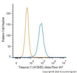
- Experimental details
- Flow Cytometry: Tenascin C Antibody (4C8MS) [NB110-68136] - An intracellular stain was performed on SK-MEL-28 cells with Tenascin C Antibody [4C8MS] NB110-68136AF647 (blue) and a matched isotype control (orange). Cells were fixed with 4% PFA and then permeabilized with 0.1% saponin. Cells were incubated in an antibody dilution of 2.5 ug/mL for 30 minutes at room temperature. Both antibodies were conjugated to Alexa Fluor 647.
