Explore
 Validate
 Learn
Learn Immunocytochemistry
Antibody data
- Antibody Data
- Antigen structure
- References [2]
- Comments [0]
- Validations
- Immunocytochemistry [1]
- Immunohistochemistry [2]
Submit
Validation data
Reference
Comment
Report error
- Product number
- GTX75897 - Provider product page

- Provider
- GeneTex
- Proper citation
- GeneTex Cat#GTX75897, RRID:AB_10728896
- Product name
- Desmoglein 2 antibody [6D8]
- Antibody type
- Monoclonal
- Host
- Mouse
- Vial size
- 100 µg
Submitted references Germline Competent Pluripotent Mouse Stem Cells Generated by Plasmid Vectors.
Measuring topology of low-intensity DNA methylation sites for high-throughput assessment of epigenetic drug-induced effects in cancer cells.
Chen CH, Su YH, Lee KH, Chuang CK
Animal biotechnology 2016;27(3):157-65
Animal biotechnology 2016;27(3):157-65
Measuring topology of low-intensity DNA methylation sites for high-throughput assessment of epigenetic drug-induced effects in cancer cells.
Gertych A, Farkas DL, Tajbakhsh J
Experimental cell research 2010 Nov 15;316(19):3150-60
Experimental cell research 2010 Nov 15;316(19):3150-60
No comments: Submit comment
Supportive validation
- Submitted by
- GeneTex (provider)
- Main image
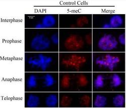
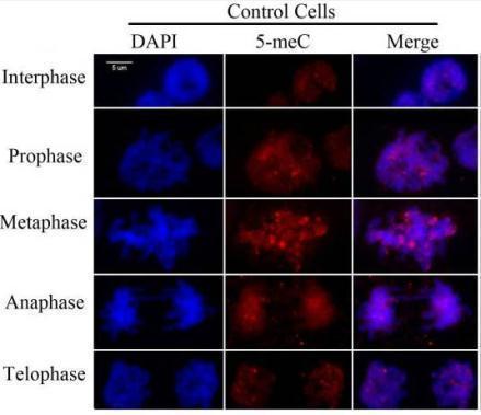
- Experimental details
- Immunostaining for 5-meC in representative maize root tip cells in interphase or mitosis using 5-meC antibody (GTX75897). Pontes, O. et al. (2007) PLoS One. 2: e1157.
Supportive validation
- Submitted by
- GeneTex (provider)
- Main image

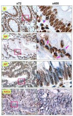
- Experimental details
- 5-MeC in DNA of various stages of colorectal lesions was detected by IHC staining using 5-MeC antibody (GTX75897). Sections of HPs, APs, AdCas and histologically ¡§normal¡¨ mucosa around colorectal cancers were stained with mAb to 5-MeC and counterstained with hematoxylin. N (> 3 ~ 4+), ¡§normal¡¨ mucosa ; HP (3+), the hyperplastic polyps; AP (2+), the adenomatous polyps; and AdCa (< 1+), adenocarcinomas. Image published in Shen et al. (2009) Int J Clin Exp Pathol. 2:21-33.
- Submitted by
- GeneTex (provider)
- Main image
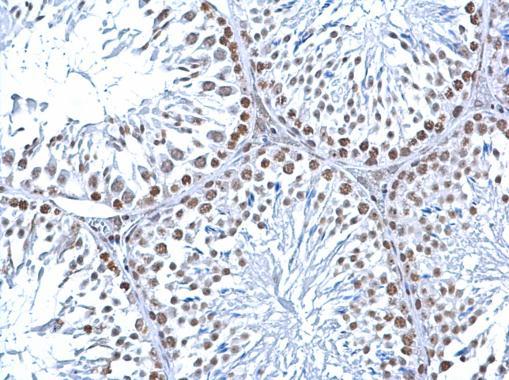
- Experimental details
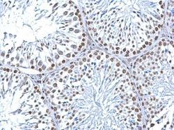
- 5-Methylcytosine / 5-mC antibody [33D3] detects 5-Methylcytosine / 5-mC at nucleus on mouse testis by immunohistochemical analysis. Sample: Paraffin-embedded mouse testis. 5-Methylcytosine / 5-mC antibody [33D3] (GTX75897) diluted at 1:200.