 Explore
 Validate
 Learn
Learn Western blot
 Immunohistochemistry
Antibody data
- Antibody Data
- Antigen structure
- References [7]
- Comments [0]
- Validations
- Western blot [3]
- Immunocytochemistry [2]
- Flow cytometry [1]
Submit
Validation data
Reference
Comment
Report error
- Product number
- MAB2055 - Provider product page

- Provider
- R&D Systems
- Product name
- Human/Mouse/Rat Akt Pan Specific Antibody
- Antibody type
- Monoclonal
- Description
- Protein A or G purified from hybridoma culture supernatant. Detects human, mouse and rat Akt in direct ELISAs and Western blots.
- Reactivity
- Human, Mouse, Rat
- Host
- Mouse
- Conjugate
- Unconjugated
- Antigen sequence
P31749- Isotype
- IgG
- Antibody clone number
- 281046
- Vial size
- 100 ug
- Concentration
- LYOPH
- Storage
- Use a manual defrost freezer and avoid repeated freeze-thaw cycles. 12 months from date of receipt, -20 to -70 °C as supplied. 1 month, 2 to 8 °C under sterile conditions after reconstitution. 6 months, -20 to -70 °C under sterile conditions after reconstitution.
Submitted references Endocrine responses and acute mTOR pathway phosphorylation to resistance exercise with leucine and whey.
Subthalamic Nucleus Deep Brain Stimulation Employs trkB Signaling for Neuroprotection and Functional Restoration.
Adipocyte SIRT1 controls systemic insulin sensitivity by modulating macrophages in adipose tissue.
Lack of CD2AP disrupts Glut4 trafficking and attenuates glucose uptake in podocytes.
Single amino acid substitutions in the chemotactic sequence of urokinase receptor modulate cell migration and invasion.
Cellular repressor of E1A-stimulated genes inhibits human vascular smooth muscle cell apoptosis via blocking P38/JNK MAP kinase activation.
Overexpressing cellular repressor of E1A-stimulated genes protects mesenchymal stem cells against hypoxia- and serum deprivation-induced apoptosis by activation of PI3K/Akt.
Lane MT, Herda TJ, Fry AC, Cooper MA, Andre MJ, Gallagher PM
Biology of sport 2017 Jun;34(2):197-203
Biology of sport 2017 Jun;34(2):197-203
Subthalamic Nucleus Deep Brain Stimulation Employs trkB Signaling for Neuroprotection and Functional Restoration.
Fischer DL, Kemp CJ, Cole-Strauss A, Polinski NK, Paumier KL, Lipton JW, Steece-Collier K, Collier TJ, Buhlinger DJ, Sortwell CE
The Journal of neuroscience : the official journal of the Society for Neuroscience 2017 Jul 12;37(28):6786-6796
The Journal of neuroscience : the official journal of the Society for Neuroscience 2017 Jul 12;37(28):6786-6796
Adipocyte SIRT1 controls systemic insulin sensitivity by modulating macrophages in adipose tissue.
Hui X, Zhang M, Gu P, Li K, Gao Y, Wu D, Wang Y, Xu A
EMBO reports 2017 Apr;18(4):645-657
EMBO reports 2017 Apr;18(4):645-657
Lack of CD2AP disrupts Glut4 trafficking and attenuates glucose uptake in podocytes.
Tolvanen TA, Dash SN, Polianskyte-Prause Z, Dumont V, Lehtonen S
Journal of cell science 2015 Dec 15;128(24):4588-600
Journal of cell science 2015 Dec 15;128(24):4588-600
Single amino acid substitutions in the chemotactic sequence of urokinase receptor modulate cell migration and invasion.
Bifulco K, Longanesi-Cattani I, Franco P, Pavone V, Mugione P, Di Carluccio G, Masucci MT, Arra C, Pirozzi G, Stoppelli MP, Carriero MV
PloS one 2012;7(9):e44806
PloS one 2012;7(9):e44806
Cellular repressor of E1A-stimulated genes inhibits human vascular smooth muscle cell apoptosis via blocking P38/JNK MAP kinase activation.
Han Y, Wu G, Deng J, Tao J, Guo L, Tian X, Kang J, Zhang X, Yan C
Journal of molecular and cellular cardiology 2010 Jun;48(6):1225-35
Journal of molecular and cellular cardiology 2010 Jun;48(6):1225-35
Overexpressing cellular repressor of E1A-stimulated genes protects mesenchymal stem cells against hypoxia- and serum deprivation-induced apoptosis by activation of PI3K/Akt.
Deng J, Han Y, Yan C, Tian X, Tao J, Kang J, Li S
Apoptosis : an international journal on programmed cell death 2010 Apr;15(4):463-73
Apoptosis : an international journal on programmed cell death 2010 Apr;15(4):463-73
No comments: Submit comment
Supportive validation
- Submitted by
- R&D Systems (provider)
- Main image
- Experimental details
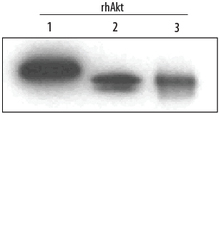
- Detection of Human Akt Pan Specific by Western Blot. Western blot shows Recombinant Human Active Akt1 (Catalog # 1775-KS), recombinant human Akt2, and recombinant human Akt3 (5 ng/lane). PVDF membrane was probed with 0.2 µg/mL Mouse Anti-Human/Mouse/Rat Akt Pan Specific Monoclonal Antibody (Catalog # MAB2055) followed by HRP-conjugated Anti-Mouse IgG Secondary Antibody (Catalog # HAF007). This experiment was conducted under reducing conditions and using Immunoblot Buffer Group 4.
- Submitted by
- R&D Systems (provider)
- Main image
- Experimental details
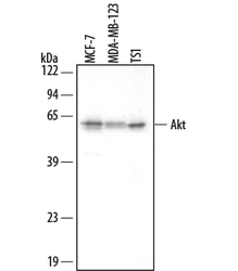
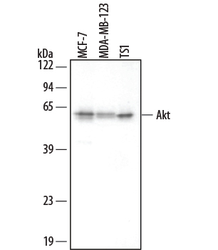
- Detection of Human/Mouse Akt by Western Blot. Western blot shows lysates of MCF-7 human breast cancer cell line, MBA-MB-123 human breast cancer cell line, and TS1 mouse helper T cell line. PVDF membrane was probed with 0.2 µg/mL Mouse Anti-Human/Mouse/Rat Akt Pan Specific Monoclonal Antibody (Catalog # MAB2055) followed by HRP-conjugated Anti-Mouse IgG Secondary Antibody (Catalog # HAF007). This experiment was conducted under reducing conditions and using Immunoblot Buffer Group 4.
- Submitted by
- R&D Systems (provider)
- Main image

- Experimental details
- Detection of Human Akt by Simple WesternTM. Simple Western lane view shows lysates of MCF-7 human breast cancer cell line, loaded at 0.2 mg/mL. A specific band was detected for Akt at approximately 62 kDa (as indicated) using 2 µg/mL of Mouse Anti-Human/Mouse/Rat Akt Pan Specific Monoclonal Antibody (Catalog # MAB2055). This experiment was conducted under reducing conditions and using the 12-230 kDa separation system.

Supportive validation
- Submitted by
- R&D Systems (provider)
- Main image

- Experimental details
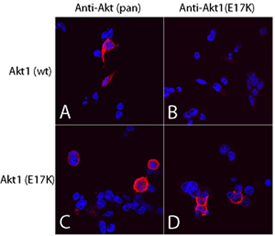
- Akt (pan) in 293T Human Cell Line. Akt pan specific (panels A and C) and Akt1 (E17K Mutation) (panels B and D) were detected in immersion fixed 293T human embryonic kidney cell line transfected with wild type (panels A and B) or E17K mutated (panels C and D) Akt1 using Mouse Anti-Human/Mouse/Rat Akt Pan Specific Monoclonal Antibody (Catalog # MAB2055) and Mouse Anti-Human Akt1 (E17K Mutation) Monoclonal Antibody (Catalog # MAB6815). Both antibodies were used at 10 µg/mL for 3 hours at room temperature. Cells were stained using the NorthernLights™ 557-conjugated Anti-Mouse IgG Secondary Antibody (red; Catalog # NL007) and counterstained with DAPI (blue). Specific staining was localized to plasma membranes and cytoplasm. View our protocol for Fluorescent ICC Staining of Cells on Coverslips.
- Submitted by
- R&D Systems (provider)
- Main image

- Experimental details
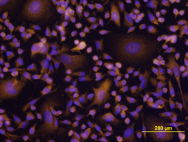
- Akt in MDA-MB-231 Human Cell Line. Akt was detected in immersion fixed MDA-MB-231 human breast cancer cell line using Mouse Anti-Human/ Mouse/Rat Akt Monoclonal Antibody (Catalog # MAB2055) at 10 µg/mL for 3 hours at room temperature. Cells were stained using the NorthernLights™ 557-conjugated Anti-Mouse IgG Secondary Antibody (yellow; Catalog # NL007) and counter-stained with DAPI (blue). View our protocol for Fluorescent ICC Staining of Cells on Coverslips.
Supportive validation
- Submitted by
- R&D Systems (provider)
- Main image
- Experimental details
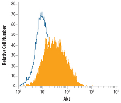
- Detection of Akt in MCF-7 Human Cell Line by Flow Cytometry. MCF-7 human breast cancer cell line was stained with Mouse Anti-Human/Mouse/Rat Akt Monoclonal Antibody (Catalog # MAB2055, filled histogram) or isotype control antibody (Catalog # MAB0041, open histogram), followed by Phycoerythrin-conjugated Anti-Mouse IgG F(ab')2 Secondary Antibody (Catalog # F0102B). To facilitate intracellular staining, cells were fixed with paraformaldehyde and permeabilized with saponin.
