 Explore
 Validate
 Learn
Learn Western blot
Antibody data
- Antibody Data
- Antigen structure
- References [22]
- Comments [0]
- Validations
- Western blot [2]
- Immunohistochemistry [1]
- Flow cytometry [1]
Submit
Validation data
Reference
Comment
Report error
- Product number
- AF887 - Provider product page

- Provider
- R&D Systems
- Product name
- Human/Mouse/Rat Phospho-Akt (S473) Pan Specific Antibody
- Antibody type
- Polyclonal
- Description
- Antigen and protein A Affinity-purified. Detects human, mouse and rat Akt1, Akt2 and Akt3, when phosphorylated at S473, S474 and S472, respectively.
- Reactivity
- Human, Mouse, Rat
- Host
- Rabbit
- Conjugate
- Unconjugated
- Isotype
- IgG
- Vial size
- 100 ug
- Concentration
- LYOPH
- Storage
- Use a manual defrost freezer and avoid repeated freeze-thaw cycles. 12 months from date of receipt, -20 to -70 °C as supplied. 1 month, 2 to 8 °C under sterile conditions after reconstitution. 6 months, -20 to -70 °C under sterile conditions after reconstitution.
Submitted references Woodchuck VEGF (wVEGF) characteristics: Model for angiogenesis and human hepatocellular carcinoma directed therapies.
Signaling Lymphocyte Activation Molecule Family 5 Enhances Autophagy and Fine-Tunes Cytokine Response in Monocyte-Derived Dendritic Cells via Stabilization of Interferon Regulatory Factor 8.
Phosphoinositide 3-kinase-delta could be a biomarker for eosinophilic nasal polyps.
Differential expression of ABCB5 in BRAF inhibitor-resistant melanoma cell lines.
Modeling multiple myeloma-bone marrow interactions and response to drugs in a 3D surrogate microenvironment.
Different dynamics of NLRP3 inflammasome-mediated IL‑1β production in GM-CSF- and M-CSF-differentiated human macrophages.
Subthalamic Nucleus Deep Brain Stimulation Employs trkB Signaling for Neuroprotection and Functional Restoration.
Dual inhibition of CDK4/Rb and PI3K/AKT/mTOR pathways by ON123300 induces synthetic lethality in mantle cell lymphomas.
Ocular Delivery of PACAP1-27 Protects the Retina From Ischemic Damage in Rodents.
Interferon-γ suppresses intestinal epithelial aquaporin-1 expression via Janus kinase and STAT3 activation.
Water-filtered infrared a irradiation in combination with visible light inhibits acute chlamydial infection.
Neuronal pentraxin 2 supports clear cell renal cell carcinoma by activating the AMPA-selective glutamate receptor-4.
The role of nuclear β-catenin accumulation in the Twist2-induced ovarian cancer EMT.
Single amino acid substitutions in the chemotactic sequence of urokinase receptor modulate cell migration and invasion.
An elaborate regulation of Mammalian target of rapamycin activity is required for somatic cell reprogramming induced by defined transcription factors.
EphB receptors trigger Akt activation and suppress Fas receptor-induced apoptosis in malignant T lymphocytes.
Cellular repressor of E1A-stimulated genes inhibits human vascular smooth muscle cell apoptosis via blocking P38/JNK MAP kinase activation.
Overexpressing cellular repressor of E1A-stimulated genes protects mesenchymal stem cells against hypoxia- and serum deprivation-induced apoptosis by activation of PI3K/Akt.
CREG inhibits migration of human vascular smooth muscle cells by mediating IGF-II endocytosis.
The involvement of the fractalkine receptor in the transmigration of neuroblastoma cells through bone-marrow endothelial cells.
Osteoblast-derived factors induce an expression signature that identifies prostate cancer metastasis and hormonal progression.
Enhancing mammalian target of rapamycin (mTOR)-targeted cancer therapy by preventing mTOR/raptor inhibition-initiated, mTOR/rictor-independent Akt activation.
Huang H, Salavaggione O, Rivera L, Mukherjee S, Brekken R, Tennant B, Iyer R, Adjei A
Archives of biochemistry and biophysics 2019 Jan;661:97-106
Archives of biochemistry and biophysics 2019 Jan;661:97-106
Signaling Lymphocyte Activation Molecule Family 5 Enhances Autophagy and Fine-Tunes Cytokine Response in Monocyte-Derived Dendritic Cells via Stabilization of Interferon Regulatory Factor 8.
Agod Z, Pazmandi K, Bencze D, Vereb G, Biro T, Szabo A, Rajnavolgyi E, Bacsi A, Engel P, Lanyi A
Frontiers in immunology 2018;9:62
Frontiers in immunology 2018;9:62
Phosphoinositide 3-kinase-delta could be a biomarker for eosinophilic nasal polyps.
Kim JS, Jeong JS, Lee KB, Kim SR, Choe YH, Kwon SH, Cho SH, Lee YC
Scientific reports 2018 Oct 30;8(1):15990
Scientific reports 2018 Oct 30;8(1):15990
Differential expression of ABCB5 in BRAF inhibitor-resistant melanoma cell lines.
Xiao J, Egger ME, McMasters KM, Hao H
BMC cancer 2018 Jun 22;18(1):675
BMC cancer 2018 Jun 22;18(1):675
Modeling multiple myeloma-bone marrow interactions and response to drugs in a 3D surrogate microenvironment.
Belloni D, Heltai S, Ponzoni M, Villa A, Vergani B, Pecciarini L, Marcatti M, Girlanda S, Tonon G, Ciceri F, Caligaris-Cappio F, Ferrarini M, Ferrero E
Haematologica 2018 Apr;103(4):707-716
Haematologica 2018 Apr;103(4):707-716
Different dynamics of NLRP3 inflammasome-mediated IL‑1β production in GM-CSF- and M-CSF-differentiated human macrophages.
Budai MM, Tőzsér J, Benkő S
Journal of leukocyte biology 2017 Jun;101(6):1335-1347
Journal of leukocyte biology 2017 Jun;101(6):1335-1347
Subthalamic Nucleus Deep Brain Stimulation Employs trkB Signaling for Neuroprotection and Functional Restoration.
Fischer DL, Kemp CJ, Cole-Strauss A, Polinski NK, Paumier KL, Lipton JW, Steece-Collier K, Collier TJ, Buhlinger DJ, Sortwell CE
The Journal of neuroscience : the official journal of the Society for Neuroscience 2017 Jul 12;37(28):6786-6796
The Journal of neuroscience : the official journal of the Society for Neuroscience 2017 Jul 12;37(28):6786-6796
Dual inhibition of CDK4/Rb and PI3K/AKT/mTOR pathways by ON123300 induces synthetic lethality in mantle cell lymphomas.
Divakar SK, Ramana Reddy MV, Cosenza SC, Baker SJ, Perumal D, Antonelli AC, Brody J, Akula B, Parekh S, Reddy EP
Leukemia 2016 Jan;30(1):86-93
Leukemia 2016 Jan;30(1):86-93
Ocular Delivery of PACAP1-27 Protects the Retina From Ischemic Damage in Rodents.
Werling D, Reglodi D, Banks WA, Salameh TS, Kovacs K, Kvarik T, Vaczy A, Kovacs L, Mayer F, Danyadi B, Lokos E, Tamas A, Toth G, Biro Z, Atlasz T
Investigative ophthalmology & visual science 2016 Dec 1;57(15):6683-6691
Investigative ophthalmology & visual science 2016 Dec 1;57(15):6683-6691
Interferon-γ suppresses intestinal epithelial aquaporin-1 expression via Janus kinase and STAT3 activation.
Dicay MS, Hirota CL, Ronaghan NJ, Peplowski MA, Zaheer RS, Carati CA, MacNaughton WK
PloS one 2015;10(3):e0118713
PloS one 2015;10(3):e0118713
Water-filtered infrared a irradiation in combination with visible light inhibits acute chlamydial infection.
Marti H, Koschwanez M, Pesch T, Blenn C, Borel N
PloS one 2014;9(7):e102239
PloS one 2014;9(7):e102239
Neuronal pentraxin 2 supports clear cell renal cell carcinoma by activating the AMPA-selective glutamate receptor-4.
von Roemeling CA, Radisky DC, Marlow LA, Cooper SJ, Grebe SK, Anastasiadis PZ, Tun HW, Copland JA
Cancer research 2014 Sep 1;74(17):4796-810
Cancer research 2014 Sep 1;74(17):4796-810
The role of nuclear β-catenin accumulation in the Twist2-induced ovarian cancer EMT.
Mao Y, Xu J, Li Z, Zhang N, Yin H, Liu Z
PloS one 2013;8(11):e78200
PloS one 2013;8(11):e78200
Single amino acid substitutions in the chemotactic sequence of urokinase receptor modulate cell migration and invasion.
Bifulco K, Longanesi-Cattani I, Franco P, Pavone V, Mugione P, Di Carluccio G, Masucci MT, Arra C, Pirozzi G, Stoppelli MP, Carriero MV
PloS one 2012;7(9):e44806
PloS one 2012;7(9):e44806
An elaborate regulation of Mammalian target of rapamycin activity is required for somatic cell reprogramming induced by defined transcription factors.
He J, Kang L, Wu T, Zhang J, Wang H, Gao H, Zhang Y, Huang B, Liu W, Kou Z, Zhang H, Gao S
Stem cells and development 2012 Sep 20;21(14):2630-41
Stem cells and development 2012 Sep 20;21(14):2630-41
EphB receptors trigger Akt activation and suppress Fas receptor-induced apoptosis in malignant T lymphocytes.
Maddigan A, Truitt L, Arsenault R, Freywald T, Allonby O, Dean J, Narendran A, Xiang J, Weng A, Napper S, Freywald A
Journal of immunology (Baltimore, Md. : 1950) 2011 Dec 1;187(11):5983-94
Journal of immunology (Baltimore, Md. : 1950) 2011 Dec 1;187(11):5983-94
Cellular repressor of E1A-stimulated genes inhibits human vascular smooth muscle cell apoptosis via blocking P38/JNK MAP kinase activation.
Han Y, Wu G, Deng J, Tao J, Guo L, Tian X, Kang J, Zhang X, Yan C
Journal of molecular and cellular cardiology 2010 Jun;48(6):1225-35
Journal of molecular and cellular cardiology 2010 Jun;48(6):1225-35
Overexpressing cellular repressor of E1A-stimulated genes protects mesenchymal stem cells against hypoxia- and serum deprivation-induced apoptosis by activation of PI3K/Akt.
Deng J, Han Y, Yan C, Tian X, Tao J, Kang J, Li S
Apoptosis : an international journal on programmed cell death 2010 Apr;15(4):463-73
Apoptosis : an international journal on programmed cell death 2010 Apr;15(4):463-73
CREG inhibits migration of human vascular smooth muscle cells by mediating IGF-II endocytosis.
Han Y, Cui J, Tao J, Guo L, Guo P, Sun M, Kang J, Zhang X, Yan C, Li S
Experimental cell research 2009 Nov 15;315(19):3301-11
Experimental cell research 2009 Nov 15;315(19):3301-11
The involvement of the fractalkine receptor in the transmigration of neuroblastoma cells through bone-marrow endothelial cells.
Nevo I, Sagi-Assif O, Meshel T, Ben-Baruch A, Jöhrer K, Greil R, Trejo LE, Kharenko O, Feinmesser M, Yron I, Witz IP
Cancer letters 2009 Jan 8;273(1):127-39
Cancer letters 2009 Jan 8;273(1):127-39
Osteoblast-derived factors induce an expression signature that identifies prostate cancer metastasis and hormonal progression.
Wang G, Haile S, Comuzzi B, Tien AH, Wang J, Yong TM, Jelescu-Bodos AE, Blaszczyk N, Vessella RL, Masri BA, Sadar MD
Cancer research 2009 Apr 15;69(8):3433-42
Cancer research 2009 Apr 15;69(8):3433-42
Enhancing mammalian target of rapamycin (mTOR)-targeted cancer therapy by preventing mTOR/raptor inhibition-initiated, mTOR/rictor-independent Akt activation.
Wang X, Yue P, Kim YA, Fu H, Khuri FR, Sun SY
Cancer research 2008 Sep 15;68(18):7409-18
Cancer research 2008 Sep 15;68(18):7409-18
No comments: Submit comment
Supportive validation
- Submitted by
- R&D Systems (provider)
- Main image

- Experimental details
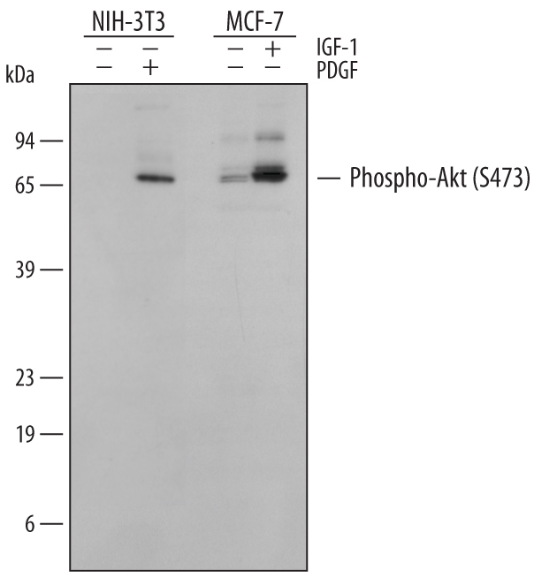
- Detection of Mouse and Human Phospho-Akt (S473) Pan Specific by Western Blot. Western blot shows lysates of NIH-3T3 mouse embryonic fibroblast cell line untreated (-) or treated (+) with 100 ng/mL Human PDGF (Catalog # 120-HD) for 15 minutes and MCF-7 human breast cancer cell line untreated or treated with 100 ng/mL Recombinant Human IGF-1 (Catalog # 291-G1) for 15 minutes. PVDF membrane was probed with 0.5 µg/mL of Rabbit Anti-Human/Mouse/Rat Phospho-Akt (S473) Pan Specific Antigen Affinity-purified Polyclonal Antibody (Catalog # AF887), followed by HRP-conjugated Anti-Rabbit IgG Secondary Antibody (Catalog # HAF008). A specific band was detected for Phospho-Akt (S473) Pan Specific at approximately 60 kDa (as indicated). This experiment was conducted under reducing conditions and using Immunoblot Buffer Group 1.
- Submitted by
- R&D Systems (provider)
- Main image
- Experimental details
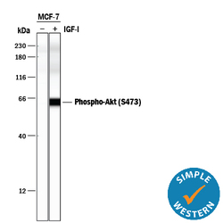
- Detection of Human Phospho-Akt (S473) by Simple WesternTM. Simple Western lane view shows lysates of MCF-7 human breast cancer cell line and A549 human lung carcinoma cell line untreated (-) or treated (+) with 100 ng/mL Recombinant Human IGF-I (Catalog # 291-G1) for 15 minutes, loaded at 0.2 mg/mL. A specific band was detected for Phospho-Akt (S473) at approximately 60 kDa (as indicated) using 5 µg/mL of Rabbit Anti-Human/Mouse/Rat Phospho-Akt (S473) Pan Specific Antigen Affinity-purified Polyclonal Antibody (Catalog # AF887). This experiment was conducted under reducing conditions and using the 12-230 kDa separation system.

Supportive validation
- Submitted by
- R&D Systems (provider)
- Main image
- Experimental details
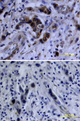
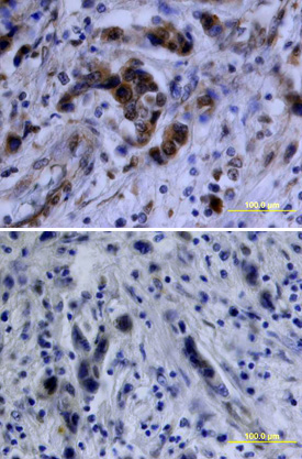
- Akt in Human Breast Cancer Tissue. Akt phosphorylated at S473 was detected in immersion fixed paraffin-embedded sections of human breast cancer tissue using Rabbit Anti-Human/Mouse/Rat Phospho-Akt (S473) Antigen Affinity-purified Polyclonal Antibody (Catalog # AF887) at 10 µg/mL overnight at 4 °C. Tissue was stained using the Anti-Rabbit HRP-DAB Cell & Tissue Staining Kit (brown; Catalog # CTS005) and counterstained with hematoxylin (blue). Lower panel shows a lack of labeling if primary antibodies are omitted and tissue is stained only with secondary antibody followed by incubation with detection reagents. View our protocol for Chromogenic IHC Staining of Paraffin-embedded Tissue Sections.
Supportive validation
- Submitted by
- R&D Systems (provider)
- Main image
- Experimental details
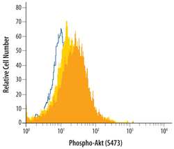
- Detection of Phospho-Akt (S473) in IGF-1-treated MCF-7 cells by Flow Cytometry. Serum-starved MCF-7 human breast cancer cells were unstimulated (light orange filled histogram) or activated with 100 ng/mL Recombinant human IGF-1 (Catalog # 291-G1, dark orange filled histogram) for 15 minutes, then stained with Rabbit Anti-Human/Mouse/Rat Phospho-Akt (S473) Antigen Affinity-purified Polyclonal Antibody (Catalog # AF887) or control antibody (Catalog # AB-105-C, open histogram), followed by Allophycocyanin-conjugated Anti-Rabbit IgG Secondary Antibody (Catalog # F0111). To facilitate intracellular staining, cells were fixed with paraformaldehyde and permeabilized with saponin.
