 Explore
 Validate
 Learn
Learn Western blot
 ELISA
ELISAAntibody data
- Antibody Data
- Antigen structure
- References [3]
- Comments [0]
- Validations
- Western blot [3]
- Immunohistochemistry [3]
Submit
Validation data
Reference
Comment
Report error
- Product number
- NB600-590 - Provider product page

- Provider
- Novus Biologicals
- Proper citation
- Novus Cat#NB600-590, RRID:AB_10002759
- Product name
- Rabbit Polyclonal AKT1 Antibody
- Antibody type
- Polyclonal
- Description
- Immunogen affinity purified. This antibody is specific for phosphorylated human AKT1 phospho S473. Minimal reactivity occurs against non-phosphorylated AKT1.
- Reactivity
- Human, Mouse, Rat
- Host
- Rabbit
- Isotype
- IgG
- Vial size
- 0.1 mg
- Concentration
- 1 mg/ml
- Storage
- Store at -20C. Avoid freeze-thaw cycles.
Submitted references Predictive microRNAs for lymph node metastasis in endoscopically resectable submucosal colorectal cancer.
GSK3β inhibition promotes synaptogenesis in Drosophila and mammalian neurons.
Temsirolimus and rituximab in patients with relapsed or refractory mantle cell lymphoma: a phase 2 study.
Jung CK, Jung SH, Yim SH, Jung JH, Choi HJ, Kang WK, Park SW, Oh ST, Kim JG, Lee SH, Chung YJ
Oncotarget 2016 May 31;7(22):32902-15
Oncotarget 2016 May 31;7(22):32902-15
GSK3β inhibition promotes synaptogenesis in Drosophila and mammalian neurons.
Cuesto G, Jordán-Álvarez S, Enriquez-Barreto L, Ferrús A, Morales M, Acebes Á
PloS one 2015;10(3):e0118475
PloS one 2015;10(3):e0118475
Temsirolimus and rituximab in patients with relapsed or refractory mantle cell lymphoma: a phase 2 study.
Ansell SM, Tang H, Kurtin PJ, Koenig PA, Inwards DJ, Shah K, Ziesmer SC, Feldman AL, Rao R, Gupta M, Erlichman C, Witzig TE
The Lancet. Oncology 2011 Apr;12(4):361-8
The Lancet. Oncology 2011 Apr;12(4):361-8
No comments: Submit comment
Supportive validation
- Submitted by
- Novus Biologicals (provider)
- Main image

- Experimental details
- Western Blot: AKT1 [p Ser473] Antibody [NB600-590] - Lane 1: AKT1 Recombinant Protein. Lane 2: AKT1 Mutant Human Recombinant Protein. Lane 3: AKT1 (phosphatase treated) Human Recombinant Protein. Load: 50 ng per lane. Primary antibody: AKT pS473 antibody at 1:1,000 for overnight at 4C. Secondary antibody: Peroxidase rabbit secondary antibody at 1:40,000 for 30 min at RT. Block: Blocking Buffer for Fluorescent Western Blotting for 30 min at RT. Predicted/Observed size: 56 kDa, 56 kDa for Gli-2. Other band(s): Gli-2 splice variants and isoforms.

- Submitted by
- Novus Biologicals (provider)
- Main image

- Experimental details
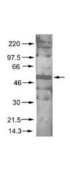
- Western Blot: AKT1 [p Ser473] Antibody [NB600-590] - The PI3K activator peptide PTD4-PI3KAc activates AKT and induces phosphorylation of GSK3 at S9.A. SH-SY5Y cell cultures were serum starved for 16 hours and challenged afterwards with PTD4-PI3KAc 21 uM, EGF 10 ng/mL or PTD4 (control peptide at a concentration of 21 uM). Cells were lysated at the indicated times and membranes incubated with anti-p-S473AKT and anti-p-S9GSK3beta. Levels of phosphorylation were normalized to AKT and GSK3beta (lower band), respectively. Image collected and cropped by CiteAb from the following publication (http://dx.plos.org/10.1371/journal.pone.0118475), licensed under a CC-BY licence.

- Submitted by
- Novus Biologicals (provider)
- Main image
- Experimental details
- Western Blot: AKT1 [p Ser473] Antibody [NB600-590] - Lane 1: nuclear extract from cells infected with adenovirus expressing nuclear-targeted AKT kinase. Load: 35 ug per lane. Primary antibody: AKT pS473 antibody at 1:200 dilution for overnight at 4C. Secondary antibody: IRDye800 rabbit secondary antibody at 1:10,000 for 45 min at RT. Block: 5% BLOTTO overnight at 4C. Predicted/Observed size: 56 kDa for AKT pS473. Other band(s): unspecific.

Supportive validation
- Submitted by
- Novus Biologicals (provider)
- Main image

- Experimental details
- Immunohistochemistry-Paraffin: AKT1 [p Ser473] Antibody [NB600-590] - Akt1 [p Ser473] Antibody [NB600-590] - Rabbit anti-AKT phospho S473 was used at 1:100 to detect AKT in human FFPE breast tumor tissue. The staining is much stronger than the weak basal level of phosphorylation in normal breast tissue. Signal was developed using Dako's Techmate streptavidin-biotin reagents.

- Submitted by
- Novus Biologicals (provider)
- Main image

- Experimental details
- Immunohistochemistry-Paraffin: AKT1 [p Ser473] Antibody [NB600-590] - Akt1 [p Ser473] Antibody [NB600-590] - Rabbit anti-AKT phospho Ser473 used at 1:100 to detect AKT in normal FFPE human breast tissue. AKT is weakly phosphorylated in normal tissue in the breast. AKt pSer473 is nuclear and occassionally cytoplasmic. Signal was developed using Dako's Techmate streptavidin-biotin reagents.

- Submitted by
- Novus Biologicals (provider)
- Main image

- Experimental details
- Immunohistochemistry-Paraffin: AKT1 [p Ser473] Antibody [NB600-590] - Akt1 [p Ser473] Antibody [NB600-590] - FFPE Human breast carcinoma tissue. Antigen retrieval: not required. AKT1 pS473 antibody at 1:100 for 1 h at RT. Secondary antibody: Dako's Techmate streptavidin-biotin reagents at 1:10,000 for 45 min at RT. Localization: Akt pS473 is nuclear and occasionally cytoplasmic.
