Explore
 Validate
 Learn
Learn Western blot
Antibody data
- Antibody Data
- Antigen structure
- References [3]
- Comments [0]
- Validations
- Western blot [1]
- Immunocytochemistry [1]
- Immunohistochemistry [1]
Submit
Validation data
Reference
Comment
Report error
- Product number
- AF4744 - Provider product page

- Provider
- R&D Systems
- Product name
- Human TBX6 Antibody
- Antibody type
- Polyclonal
- Description
- Antigen Affinity-purified. Detects human TBX6 in direct ELISAs and Western blots.
- Reactivity
- Human
- Host
- Goat
- Conjugate
- Unconjugated
- Antigen sequence
O95947- Isotype
- IgG
- Vial size
- 100 ug
- Concentration
- LYOPH
- Storage
- Use a manual defrost freezer and avoid repeated freeze-thaw cycles. 12 months from date of receipt, -20 to -70 °C as supplied. 1 month, 2 to 8 °C under sterile conditions after reconstitution. 6 months, -20 to -70 °C under sterile conditions after reconstitution.
Submitted references Lineage tracing of neuromesodermal progenitors reveals novel Wnt-dependent roles in trunk progenitor cell maintenance and differentiation.
In vitro generation of neuromesodermal progenitors reveals distinct roles for wnt signalling in the specification of spinal cord and paraxial mesoderm identity.
Generation of human vascular smooth muscle subtypes provides insight into embryological origin-dependent disease susceptibility.
Garriock RJ, Chalamalasetty RB, Kennedy MW, Canizales LC, Lewandoski M, Yamaguchi TP
Development (Cambridge, England) 2015 May 1;142(9):1628-38
Development (Cambridge, England) 2015 May 1;142(9):1628-38
In vitro generation of neuromesodermal progenitors reveals distinct roles for wnt signalling in the specification of spinal cord and paraxial mesoderm identity.
Gouti M, Tsakiridis A, Wymeersch FJ, Huang Y, Kleinjung J, Wilson V, Briscoe J
PLoS biology 2014 Aug;12(8):e1001937
PLoS biology 2014 Aug;12(8):e1001937
Generation of human vascular smooth muscle subtypes provides insight into embryological origin-dependent disease susceptibility.
Cheung C, Bernardo AS, Trotter MW, Pedersen RA, Sinha S
Nature biotechnology 2012 Jan 15;30(2):165-73
Nature biotechnology 2012 Jan 15;30(2):165-73
No comments: Submit comment
Supportive validation
- Submitted by
- R&D Systems (provider)
- Main image
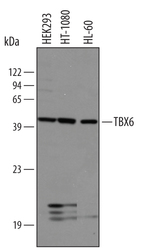
- Experimental details
- Detection of Human TBX6 by Western Blot. Western blot shows lysates of HEK293 human embryonic kidney cell line, HT1080 human fibrosarcoma cell line, and HL-60 human acute promyelocytic leukemia cell line. PVDF membrane was probed with 1 µg/mL of Goat Anti-Human TBX6 Antigen Affinity-purified Polyclonal Antibody (Catalog # AF4744) followed by HRP-conjugated Anti-Goat IgG Secondary Antibody (Catalog # HAF019). A specific band was detected for TBX6 at approximately 45 kDa (as indicated). This experiment was conducted under reducing conditions and using Immunoblot Buffer Group 8.

Supportive validation
- Submitted by
- R&D Systems (provider)
- Main image
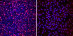
- Experimental details
- TBX6 in JOY6 Human iPS Cells. TBX6 was detected in immersion fixed JOY6 human induced pluripotent stem cells undifferentiated (right panel) and differentiated into mesoderm (left panel) using Goat Anti-Human TBX6 Antigen Affinity-purified Polyclonal Antibody (Catalog # AF4744) at 0.2 µg/mL for 3 hours at room temperature. Cells were stained using the NorthernLights™ 557-conjugated Anti-Goat IgG Secondary Antibody (red; Catalog # NL001) and counterstained with DAPI (blue). Specific staining was localized to nuclei in mesoderm. View our protocol for Fluorescent ICC Staining of Stem Cells on Coverslips.

Supportive validation
- Submitted by
- R&D Systems (provider)
- Main image
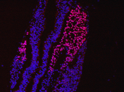
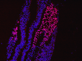
- Experimental details
- TBX6 in Embryonic Mouse Mesoderm. TBX6 was detected in immersion fixed frozen sections of embryonic mouse mesoderm (E9.5) using 10 µg/mL Human TBX6 Antigen Affinity-purified Polyclonal Antibody (Catalog # AF4744) overnight at 4 °C. Tissue was stained with the NorthernLights™ 557-conjugated Anti-Goat IgG Secondary Antibody (red; Catalog # NL001) and counterstained with DAPI (blue). View our protocol for Fluorescent IHC Staining of Frozen Tissue Sections.