 Explore
 Validate
 Learn
Learn Western blot
Antibody data
- Antibody Data
- Antigen structure
- References [2]
- Comments [0]
- Validations
- Western blot [2]
- Immunocytochemistry [2]
- Immunohistochemistry [1]
Submit
Validation data
Reference
Comment
Report error
- Product number
- GTX102637 - Provider product page

- Provider
- GeneTex
- Proper citation
- GeneTex Cat#GTX102637, RRID:AB_1949561
- Product name
- ACAT1 antibody [N1N3]
- Antibody type
- Polyclonal
- Reactivity
- Human, Mouse
- Host
- Rabbit
Submitted references Mitochondrial proteomics with siRNA knockdown to reveal ACAT1 and MDH2 in the development of doxorubicin-resistant uterine cancer.
The inositol 5-phosphatase SHIP2 is an effector of RhoA and is involved in cell polarity and migration.
Lo YW, Lin ST, Chang SJ, Chan CH, Lyu KW, Chang JF, May EW, Lin DY, Chou HC, Chan HL
Journal of cellular and molecular medicine 2015 Apr;19(4):744-59
Journal of cellular and molecular medicine 2015 Apr;19(4):744-59
The inositol 5-phosphatase SHIP2 is an effector of RhoA and is involved in cell polarity and migration.
Kato K, Yazawa T, Taki K, Mori K, Wang S, Nishioka T, Hamaguchi T, Itoh T, Takenawa T, Kataoka C, Matsuura Y, Amano M, Murohara T, Kaibuchi K
Molecular biology of the cell 2012 Jul;23(13):2593-604
Molecular biology of the cell 2012 Jul;23(13):2593-604
No comments: Submit comment
Supportive validation
- Submitted by
- GeneTex (provider)
- Main image
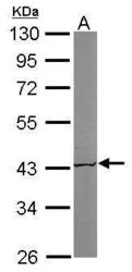
- Experimental details
- Sample (50 ug of whole cell lysate) A: Mouse brain 10% SDS PAGE GTX102637 diluted at 1:1000

- Submitted by
- GeneTex (provider)
- Main image

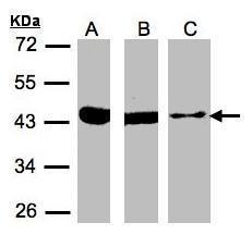
- Experimental details
- Sample(30 £gg of whole cell lysate)A:293TB:A431(GTX27909)C:MOLT4(GTX27912)10% SDS PAGEGTX102637 diluted at 1:500
Supportive validation
- Submitted by
- GeneTex (provider)
- Main image

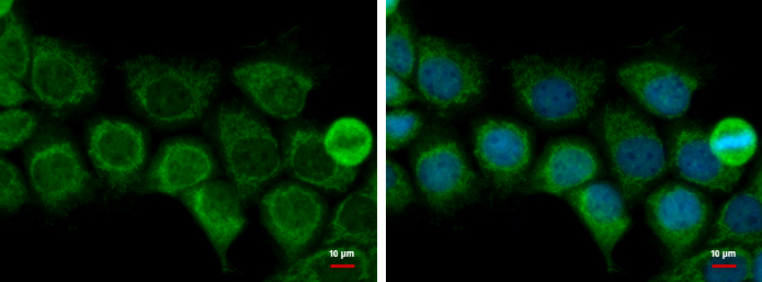
- Experimental details
- ACAT1 antibody [N1N3] detects ACAT1 protein at mitochondria by immunofluorescent analysis.Sample: A431 cells were fixed in 2% paraformaldehyde/culture medium at 37¢J for 30 min.Green: ACAT1 protein stained by ACAT1 antibody [N1N3] (GTX102637) diluted at 1:500.Blue: Hoechst 33342 staining.
- Submitted by
- GeneTex (provider)
- Main image
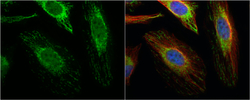
- Experimental details
- ACAT1 antibody [N1N3] detects ACAT1 protein at mitochondria by immunofluorescent analysis.Sample: HeLa cells were fixed in 4% paraformaldehyde at RT for 15 min.Green: ACAT1 protein stained by ACAT1 antibody [N1N3] (GTX102637) diluted at 1:500.Red: alpha Tubulin, a cytoskeleton marker, stained by alpha Tubulin antibody [B-5-1-2] (GTX11304) diluted at 1:10000.Blue: Hoechst 33342 staining.

Supportive validation
- Submitted by
- GeneTex (provider)
- Main image

- Experimental details
- ACAT1 antibody [N1N3] detects ACAT1 protein at cytoplasm on human normal kidney by immunohistochemical analysis. Sample: Paraffin-embedded human normal kidney. ACAT1 antibody [N1N3] (GTX102637) diluted at 1:500.
