Explore
 Validate
 Learn
Learn Western blot
 Immunocytochemistry
Antibody data
- Antibody Data
- Antigen structure
- References [2]
- Comments [0]
- Validations
- Western blot [2]
- Immunocytochemistry [1]
- Immunohistochemistry [6]
Submit
Validation data
Reference
Comment
Report error
- Product number
- HPA004428 - Provider product page

- Provider
- Atlas Antibodies
- Proper citation
- Atlas Antibodies Cat#HPA004428, RRID:AB_1078088
- Product name
- Anti-ACAT1
- Antibody type
- Polyclonal
- Reactivity
- Human, Mouse, Rat
- Host
- Rabbit
- Conjugate
- Unconjugated
- Antigen sequence
VSATRTPIGSFLGSLSLLPATKLGSIAIQGAIEKA
GIPKEEVKEAYMGNVLQGGEGQAPTRQAVLGAGLP
ISTPCTTINKVCASGMKAIMMASQSLMCGHQDVMV
AGGMESMSNVPYVMNRGSTPYGGVKLEDLIVKDGL
TD- Isotype
- IgG
- Vial size
- 100 µl
- Storage
- Store at +4°C for short term storage. Long time storage is recommended at -20°C.
Submitted references Ethanol exposure induces the cancer-associated fibroblast phenotype and lethal tumor metabolism: implications for breast cancer prevention.
Systematic validation of antibody binding and protein subcellular localization using siRNA and confocal microscopy
Sanchez-Alvarez R, Martinez-Outschoorn UE, Lin Z, Lamb R, Hulit J, Howell A, Sotgia F, Rubin E, Lisanti MP
Cell cycle (Georgetown, Tex.) 2013 Jan 15;12(2):289-301
Cell cycle (Georgetown, Tex.) 2013 Jan 15;12(2):289-301
Systematic validation of antibody binding and protein subcellular localization using siRNA and confocal microscopy
Stadler C, Hjelmare M, Neumann B, Jonasson K, Pepperkok R, Uhlén M, Lundberg E
Journal of Proteomics 2012 April;75(7):2236-2251
Journal of Proteomics 2012 April;75(7):2236-2251
No comments: Submit comment
Enhanced validation
- Submitted by
- Atlas Antibodies (provider)
- Enhanced method
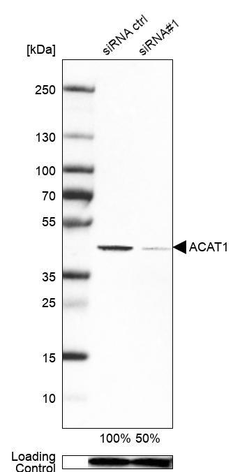
- Genetic validation
- Main image

- Experimental details
- Western blot analysis in Caco-2 cells transfected with control siRNA, target specific siRNA probe #1, using Anti-ACAT1 antibody. Remaining relative intensity is presented. Loading control: Anti-PPIB.
- Submitted by
- Atlas Antibodies (provider)
- Enhanced method
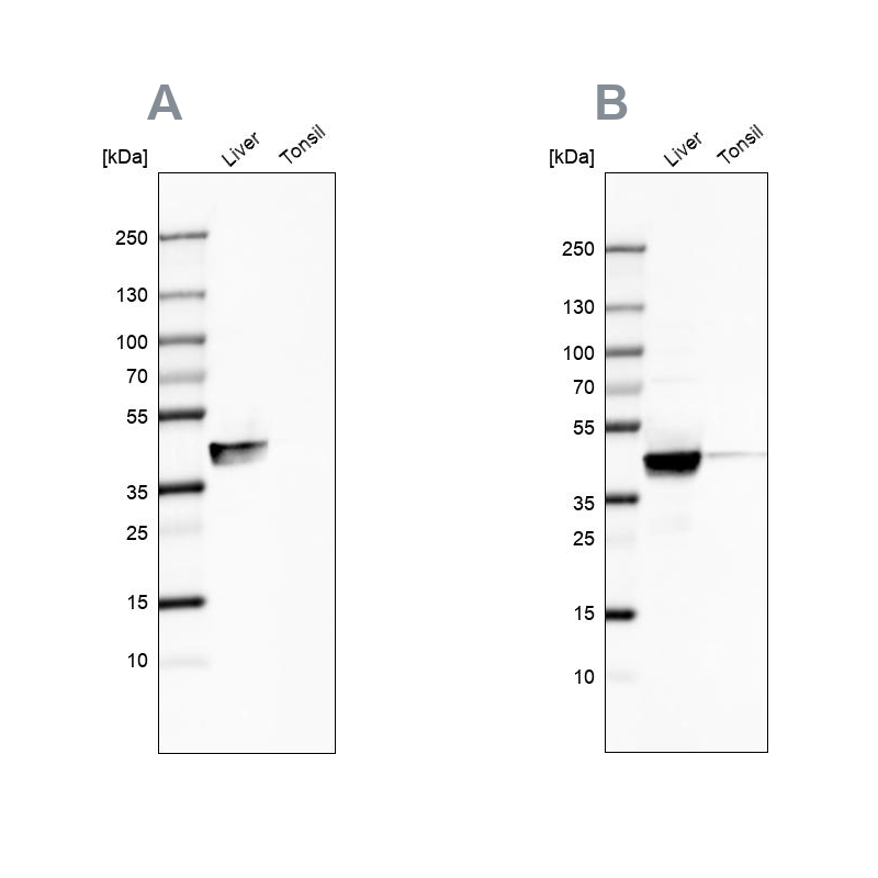
- Independent antibody validation
- Main image
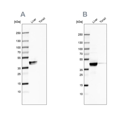
- Experimental details
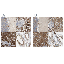
- Western blot analysis using Anti-ACAT1 antibody HPA004428 (A) shows similar pattern to independent antibody HPA007569 (B).
Supportive validation
- Submitted by
- Atlas Antibodies (provider)
- Main image
- Experimental details
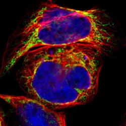
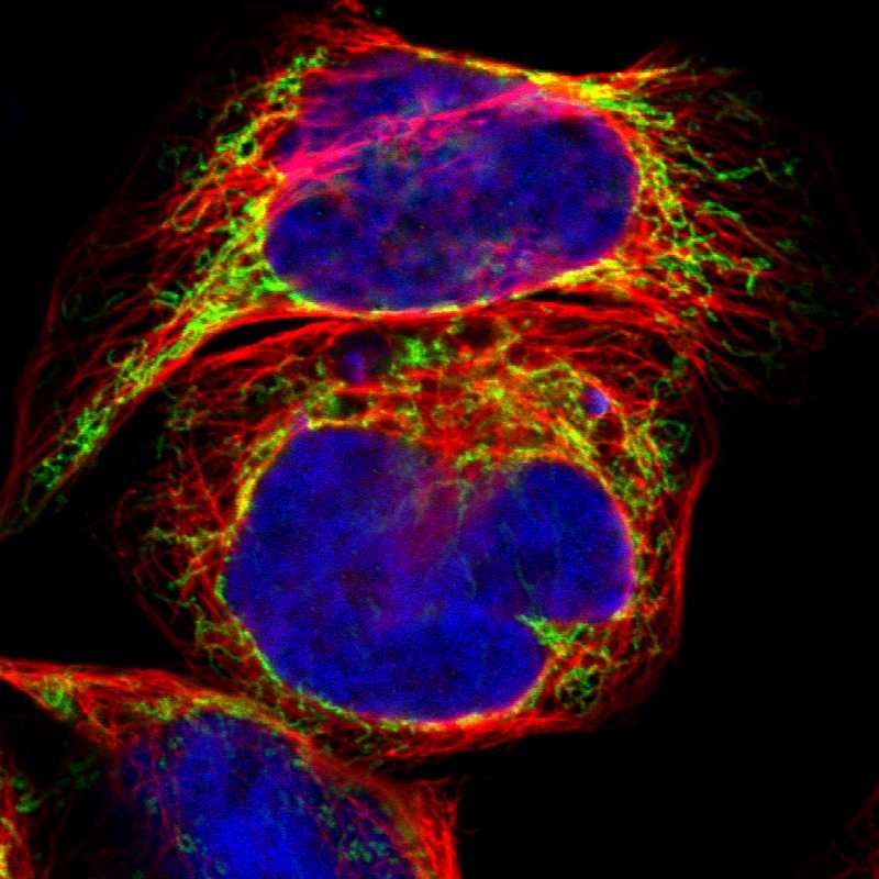
- Immunofluorescent staining of human cell line A-431 shows localization to mitochondria.
- Sample type
- HUMAN
Enhanced validation
Supportive validation
- Submitted by
- Atlas Antibodies (provider)
- Enhanced method
- Independent antibody validation
- Main image
- Experimental details
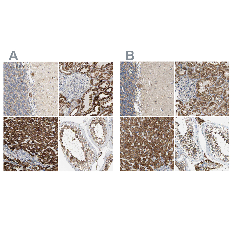
- Immunohistochemical staining of human cerebellum, kidney, liver and testis using Anti-ACAT1 antibody HPA004428 (A) shows similar protein distribution across tissues to independent antibody HPA007569 (B).
Supportive validation
- Submitted by
- Atlas Antibodies (provider)
- Main image
- Experimental details
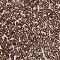
- Immunohistochemical staining of human liver shows strong cytoplasmic positivity in hepatocytes.
- Submitted by
- Atlas Antibodies (provider)
- Main image
- Experimental details
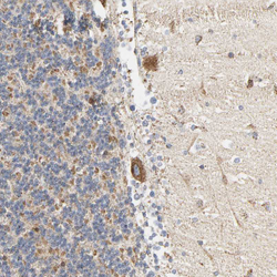
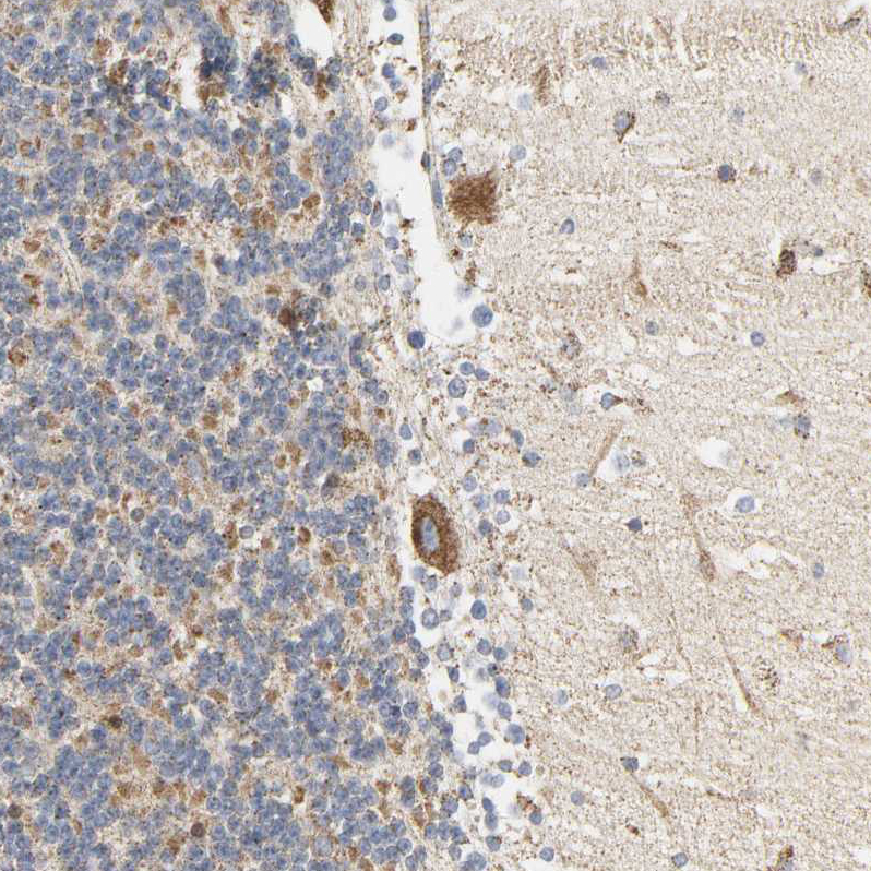
- Immunohistochemical staining of human cerebellum shows strong positivity in mitochondria in purkinje cells.
- Sample type
- HUMAN
- Submitted by
- Atlas Antibodies (provider)
- Main image
- Experimental details
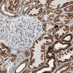
- Immunohistochemical staining of human kidney shows strong positivity in mitochondria in cells in tubules.
- Sample type
- HUMAN

- Submitted by
- Atlas Antibodies (provider)
- Main image

- Experimental details
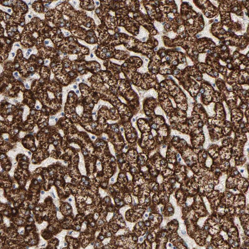
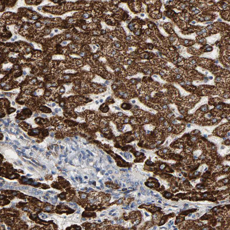
- Immunohistochemical staining of human liver shows strong positivity in mitochondria in hepatocytes.
- Sample type
- HUMAN
- Submitted by
- Atlas Antibodies (provider)
- Main image
- Experimental details
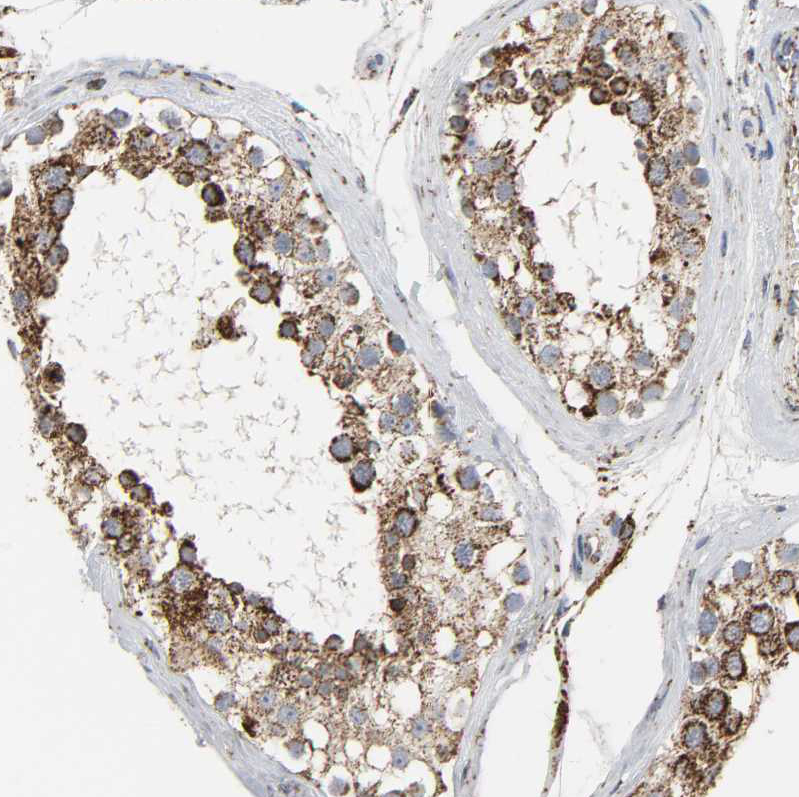
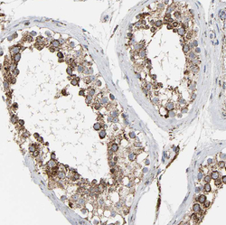
- Immunohistochemical staining of human testis shows strong positivity in mitochondria in cells in seminiferous ducts.
- Sample type
- HUMAN