Explore
 Validate
 Learn
Learn Western blot
 Immunocytochemistry
Antibody data
- Antibody Data
- Antigen structure
- References [5]
- Comments [0]
- Validations
- Western blot [1]
- Immunocytochemistry [1]
- Immunohistochemistry [6]
Submit
Validation data
Reference
Comment
Report error
- Product number
- HPA007569 - Provider product page

- Provider
- Atlas Antibodies
- Proper citation
- Atlas Antibodies Cat#HPA007569, RRID:AB_1844482
- Product name
- Anti-ACAT1
- Antibody type
- Polyclonal
- Reactivity
- Human
- Host
- Rabbit
- Conjugate
- Unconjugated
- Antigen sequence
NEQDAYAINSYTRSKAAWEAGKFGNEVIPVTVTVK
GQPDVVVKEDEEYKRVDFSKVPKLKTVFQKENGTV
TAANASTLNDGAAALVLMTADAAKRLNVTPLARIV
AFADAAVEPIDFPIAPVYAASMV- Isotype
- IgG
- Vial size
- 100 µl
- Storage
- Store at +4°C for short term storage. Long time storage is recommended at -20°C.
Submitted references Ketone bodies and two-compartment tumor metabolism: Stromal ketone production fuels mitochondrial biogenesis in epithelial cancer cells
Antibodies Biotinylated Using a Synthetic Z-domain from Protein A Provide Stringent In Situ Protein Detection
Ketolytic and glycolytic enzymatic expression profiles in malignant gliomas: implication for ketogenic diet therapy
Ketone body utilization drives tumor growth and metastasis.
Systematic validation of antibody binding and protein subcellular localization using siRNA and confocal microscopy
Martinez-Outschoorn U, Lin Z, Whitaker-Menezes D, Howell A, Lisanti M, Sotgia F
Cell Cycle 2014 November;11(21):3956-3963
Cell Cycle 2014 November;11(21):3956-3963
Antibodies Biotinylated Using a Synthetic Z-domain from Protein A Provide Stringent In Situ Protein Detection
Andersson S, Konrad A, Ashok N, Ponten F, Hober S, Asplund A
Journal of Histochemistry & Cytochemistry 2013 October;61(11):773-784
Journal of Histochemistry & Cytochemistry 2013 October;61(11):773-784
Ketolytic and glycolytic enzymatic expression profiles in malignant gliomas: implication for ketogenic diet therapy
Chang H, Olson L, Schwartz K
Nutrition & Metabolism 2013 ;10(1):47
Nutrition & Metabolism 2013 ;10(1):47
Ketone body utilization drives tumor growth and metastasis.
Martinez-Outschoorn UE, Lin Z, Whitaker-Menezes D, Howell A, Sotgia F, Lisanti MP
Cell cycle (Georgetown, Tex.) 2012 Nov 1;11(21):3964-71
Cell cycle (Georgetown, Tex.) 2012 Nov 1;11(21):3964-71
Systematic validation of antibody binding and protein subcellular localization using siRNA and confocal microscopy
Stadler C, Hjelmare M, Neumann B, Jonasson K, Pepperkok R, Uhlén M, Lundberg E
Journal of Proteomics 2012 April;75(7):2236-2251
Journal of Proteomics 2012 April;75(7):2236-2251
No comments: Submit comment
Enhanced validation
- Submitted by
- Atlas Antibodies (provider)
- Enhanced method
- Genetic validation
- Main image
- Experimental details
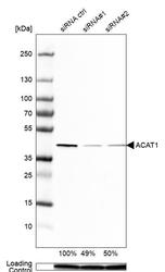
- Western blot analysis in Caco-2 cells transfected with control siRNA, target specific siRNA probe #1 and #2, using Anti-ACAT1 antibody. Remaining relative intensity is presented. Loading control: Anti-PPIB.

Supportive validation
- Submitted by
- Atlas Antibodies (provider)
- Main image
- Experimental details
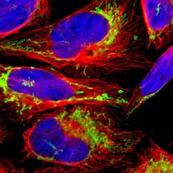
- Immunofluorescent staining of human cell line U-2 OS shows localization to mitochondria.
- Sample type
- HUMAN
Enhanced validation
Supportive validation
- Submitted by
- Atlas Antibodies (provider)
- Enhanced method
- Independent antibody validation
- Main image
- Experimental details
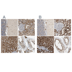
- Immunohistochemical staining of human cerebellum, kidney, liver and testis using Anti-ACAT1 antibody HPA007569 (A) shows similar protein distribution across tissues to independent antibody HPA004428 (B).

Supportive validation
- Submitted by
- Atlas Antibodies (provider)
- Main image
- Experimental details
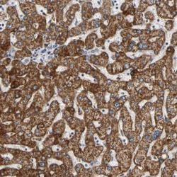
- Immunohistochemical staining of human liver shows strong cytoplasmic positivity in hepatocytes.

- Submitted by
- Atlas Antibodies (provider)
- Main image
- Experimental details
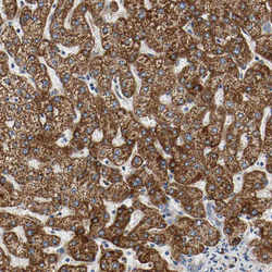
- Immunohistochemical staining of human liver shows strong positivity in mitochondria in hepatocytes.
- Sample type
- HUMAN

- Submitted by
- Atlas Antibodies (provider)
- Main image
- Experimental details
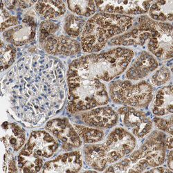
- Immunohistochemical staining of human kidney shows strong positivity in mitochondria in cells in tubules.
- Sample type
- HUMAN

- Submitted by
- Atlas Antibodies (provider)
- Main image
- Experimental details
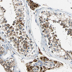
- Immunohistochemical staining of human testis shows strong positivity in mitochondria in cells in seminiferous ducts.
- Sample type
- HUMAN

- Submitted by
- Atlas Antibodies (provider)
- Main image
- Experimental details
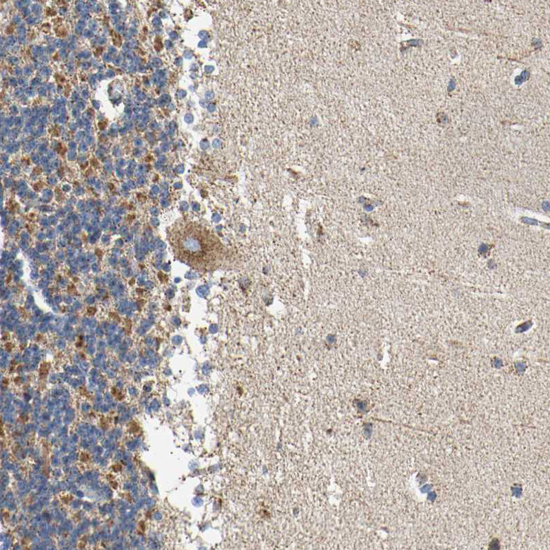
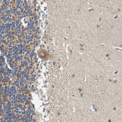
- Immunohistochemical staining of human cerebellum shows moderate positivity in mitochondria in Purkinje cells.
- Sample type
- HUMAN