 Explore
 Validate
 Learn
Learn Western blot
Antibody data
- Antibody Data
- Antigen structure
- References [0]
- Comments [0]
- Validations
- Western blot [3]
- Immunocytochemistry [1]
- Immunohistochemistry [1]
Submit
Validation data
Reference
Comment
Report error
- Product number
- MA5-45977 - Provider product page

- Provider
- Invitrogen Antibodies
- Product name
- DEPDC6 Recombinant Rabbit Monoclonal Antibody (PH00-03)
- Antibody type
- Monoclonal
- Antigen
- Recombinant full-length protein
- Description
- Predicted band size: 46 kDa Positive Control: MCF-7 cell lysate, Daudi cell lysate, PC-3M cell lysate, N2A cell lysate, L6 cell lysate, mouse brain tissue lysate, human small intestine tissue. Sequence Similarities: 96.35%Mouse; 97.73% Rat. Tissue Specificity: Expression is negatively regulated by both mTORC1 and mTORC2 (at protein level).
- Reactivity
- Human, Mouse, Rat
- Host
- Rabbit
- Isotype
- IgG
- Antibody clone number
- PH00-03
- Vial size
- 100 µL
- Concentration
- 1 mg/mL
- Storage
- Store at 4°C short term. For long term storage, store at -20°C, avoiding freeze/thaw cycles.
No comments: Submit comment
Supportive validation
- Submitted by
- Invitrogen Antibodies (provider)
- Main image

- Experimental details
- Western blot was performed using DEPDC6 Recombinant Rabbit Monoclonal Antibody (PH00-03) (Product # MA5-45977), and a 46 kDa band corresponding to DEPDC6 was observed across the cell lines tested. Whole-cell extracts of MCF-7 (20 µg lysate) (Lane 1), Daudi (10 µg lysate) (Lane 2), PC-3M (17 µg lysate) (Lane 3), N2A (15 µg lysate) (Lane 4), L6 (13 µg lysate) (Lane 5), and mouse brain tissue (20 µg lysate) (Lane 6) were electrophoresed using 12% SDS-PAGE gel. Resolved proteins were transferred onto a PVDF membrane. The blot was blocked with 5% NFDM/TBST for one hour at room temperature, then probed with the primary antibody (1:1,000 dilution) for 2 hours at room temperature and detected by chemiluminescence with HRP labeled Goat anti-Rabbit IgG secondary antibody.

- Submitted by
- Invitrogen Antibodies (provider)
- Main image

- Experimental details
- Western blot was performed using DEPDC6 Recombinant Rabbit Monoclonal Antibody (PH00-03) (Product # MA5-45977) and a 42 kDa band corresponding to DEPDC6 was observed across cell lines tested. Whole cell extracts (30 µg lysate) of HeLa (Lane 1), MCF7 (Lane 2), THP-1 (Lane 3) and Neuro-2a (Lane 4) were electrophoresed using NuPAGE™ 4-12% Bis-Tris Protein Gel (Product # NP0322BOX). Resolved proteins were then transferred onto a nitrocellulose membrane (Product # IB23002) by iBlot® 2 Dry Blotting System (Product # IB21001). The blot was probed with the primary antibody (1:1,000 dilution) and detected by chemiluminescence with Goat anti-Rabbit IgG (H+L) Superclonal™ Recombinant Secondary Antibody, HRP (Product # A27036, 1:20,000 dilution) using the iBright™ FL1500 Imaging System (Product # A44115). Chemiluminescent detection was performed using SuperSignal™ West Pico PLUS Chemiluminescent Substrate (Product # 34580).

- Submitted by
- Invitrogen Antibodies (provider)
- Main image

- Experimental details
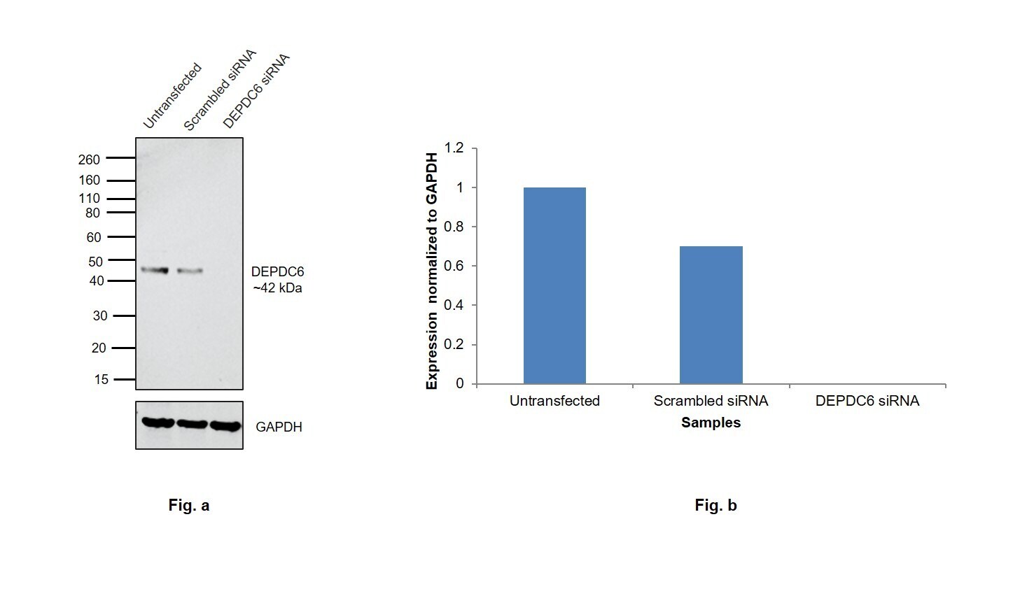
- Knockdown of DEPDC6 was achieved by transfecting HeLa with DEPDC6 specific siRNAs (Silencer® select Product # s34968 and s34970). Western blot analysis (Fig. a) was performed using Whole cell extracts from the untransfected cells (lane 1), non-targeting scrambled siRNA transfected cells (lane 2), and DEPDC6 knockdown cells (lane 3). The blot was probed with DEPDC6 Recombinant Rabbit Monoclonal Antibody (PH00-03) (Product # MA5-45977, 1:1,000 dilution) and Goat anti-Rabbit IgG (H+L) Superclonal™ Recombinant Secondary Antibody, HRP (Product # A27036, 1:20,,000 dilution). Densitometric analysis of this western blot is shown in histogram (Fig. b). Decrease in signal upon siRNA mediated knock down confirms that antibody is specific to DEPDC6.
Supportive validation
- Submitted by
- Invitrogen Antibodies (provider)
- Main image

- Experimental details
- Immunofluorescence analysis of DEPDC6 was performed using 70% confluent log phase HeLa cells. The cells were fixed with 4% paraformaldehyde for 10 minutes, permeabilized with 0.1% Triton™ X-100 for 15 minutes, and blocked with 2% BSA for 1 hour at room temperature. The cells were labeled with DEPDC6 Recombinant Rabbit Monoclonal Antibody (PH00-03) (Product # MA5-45977) at 1:100 dilution in 0.1% BSA, incubated at 4 degree celsius overnight and then labeled with Donkey anti-Rabbit IgG (H+L) Highly Cross-Adsorbed Secondary Antibody, Alexa Fluor Plus 488 (Product # A32790), 1:2,000 dilution, for 45 minutes at room temperature (Panel a: Green). Nuclei (Panel b:Blue) were stained with ProLong™ Diamond Antifade Mountant with DAPI (Product # P36962). F-actin (Panel c: Red) was stained with Rhodamine Phalloidin (Product # R415, 1:300). Panel d represents the merged image showing cytoplasmic localization. Panel e represents control cells with no primary antibody to assess background. The images were captured at 60X magnification.

Supportive validation
- Submitted by
- Invitrogen Antibodies (provider)
- Main image

- Experimental details
- Immunohistochemical analysis of DEPDC6 on formalin-fixed paraffin-embedded human small intestine tissue. The section was pre-treated using heat mediated antigen retrieval with Tris-EDTA buffer (pH 9.0) for 20 minutes. The tissue was blocked with1% BSA for 20 minutes at room temperature, then probed with DEPDC6 Recombinant Rabbit Monoclonal Antibody (PH00-03) (Product # MA5-45977) at 1:200 dilution for 60 minutes at room temperature. HRP conjugated compact polymer system and DAB chromogen were used as the detection system, followed by counterstaining with hematoxylin. The slide was mounted with DPX and the image was captured at 60X magnification.
