 Explore
 Validate
 Learn
Learn Western blot
 Immunoprecipitation
Antibody data
- Antibody Data
- Antigen structure
- References [0]
- Comments [0]
- Validations
- Western blot [3]
- Immunocytochemistry [1]
- Immunohistochemistry [1]
- Other assay [1]
Submit
Validation data
Reference
Comment
Report error
- Product number
- PA5-112261 - Provider product page

- Provider
- Invitrogen Antibodies
- Product name
- ZNF313 Polyclonal Antibody
- Antibody type
- Polyclonal
- Antigen
- Other
- Reactivity
- Human, Mouse
- Host
- Rabbit
- Isotype
- IgG
- Vial size
- 100 µL
- Concentration
- 1 mg/mL
- Storage
- Store at 4°C short term. For long term storage, store at -20°C, avoiding freeze/thaw cycles.
No comments: Submit comment
Supportive validation
- Submitted by
- Invitrogen Antibodies (provider)
- Main image
- Experimental details
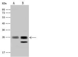
- Western Blot analysis of ZNF313 using ZNF313 Polyclonal Antibody (Product # PA5-112261) at a dilution of 1:500. Lane A: HepG2 whole cell lysate, Lane B: 293T whole cell lysate, (Lysates/proteins at 30 µg per lane). The secondary antibody used was a Goat Anti-Rabbit IgG (H+L)/HRP at 1:10,000 dilution. Developed using the ECL technique and performed under reducing conditions. Predicted band size: 26 kDa. Observed band size: 26 kDa.

- Submitted by
- Invitrogen Antibodies (provider)
- Main image
- Experimental details
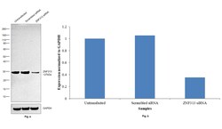
- Knockdown of ZNF313 was achieved by transfecting Jurkat with ZNF313 specific siRNAs (Silencer® select Product # S31751, S31752). Western blot analysis (Fig. a) was performed using whole cell extracts from the ZNF313 knockdown cells (lane 3), non-targeting scrambled siRNA transfected cells (lane 2) and untransfected cells (lane 1). The blot was probed with ZNF313 Polyclonal Antibody (Product # PA5-112261, 1:2000) and Goat anti-Rabbit IgG (H+L) Superclonal™ Recombinant Secondary Antibody, HRP (Product # A27036, 1:20,000) and detected by chemiluminescence using the iBright™ FL1500 Imaging System (Product # A44115). Densitometric analysis of this western blot is shown in histogram (Fig. b). Decrease in signal upon siRNA mediated knock down confirms that antibody is specific to ZNF313.

- Submitted by
- Invitrogen Antibodies (provider)
- Main image
- Experimental details
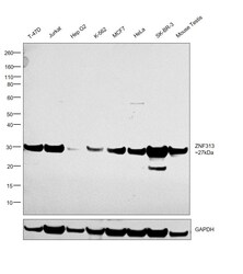
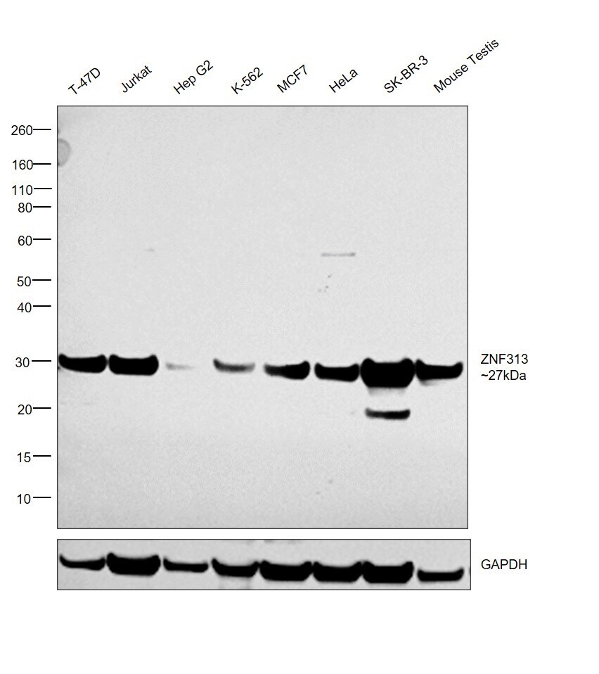
- Western blot was performed using ZNF313 Polyclonal Antibody (Product # PA5-112261) and a 27 kDa band corresponding to ZNF313 was observed across all cell lines and tissues tested. Whole cell extracts (30 µg lysate) of T-47D (Lane 1), Jurkat (Lane 2), Hep G2 (Lane 3), K-562 (Lane 4), MCF7 (Lane 5), HeLa (Lane 6), SK-BR-3 (Lane 7) and Mouse Testis (Lane 8) were electrophoresed using NuPAGE™ 4-12% Bis-Tris Protein Gel (Product # NP0321BOX), 10 well. Resolved proteins were then transferred onto a nitrocellulose membrane (Product # IB23001) by iBlot® 2 Dry Blotting System (Product # IB21001). The blot was probed with the primary antibody (1:2000) and detected by chemiluminescence with Goat anti-Rabbit IgG (H+L) Superclonal™ Recombinant Secondary Antibody, HRP (Product # A27036, 1:20,000) using the iBright™ FL1500 Imaging System (Product # A44115). Chemiluminescent detection was performed using SuperSignal™ West Pico PLUS Chemiluminescent Substrate (Product # 34580).
Supportive validation
- Submitted by
- Invitrogen Antibodies (provider)
- Main image
- Experimental details
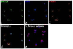
- Immunofluorescence analysis of ZNF313 was performed using 70% confluent log phase Jurkat cells. The cells were fixed with 4% paraformaldehyde for 10 minutes, permeabilized with 0.1% Triton™ X-100 for 15 minutes, and blocked with 2% BSA for 1 hour at room temperature. The cells were labeled with ZNF313 Polyclonal Antibody (Product # PA5-112261, 1:250) in 0.1% BSA, incubated at 4 degree celsius overnight and then labeled with Goat anti-Rabbit IgG (Heavy chain), Superclonal™ Recombinant Secondary Antibody, Alexa Fluor™ 488 (Product # A27034, 1:2000) for 45 minutes at room temperature (Panel a: Green). Nuclei (Panel b:Blue) were stained with ProLong™ Diamond Antifade Mountant with DAPI (Product # P36962). F-actin (Panel c: Red) was stained with Rhodamine Phalloidin (Product # R415, 1:300). Panel d represents the merged image showing cytoplasmic localization. Panel e represents control cells with no primary antibody to assess background. The images were captured at 60x magnification.

Supportive validation
- Submitted by
- Invitrogen Antibodies (provider)
- Main image
- Experimental details
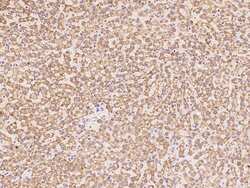
- Immunohistochemistry analysis of ZNF313 in formalin-fixed paraffin embedded sections human liver tissue sections using ZNF313 Polyclonal Antibody (Product # PA5-112261) at a dilution of 1:100.

Supportive validation
- Submitted by
- Invitrogen Antibodies (provider)
- Main image
- Experimental details
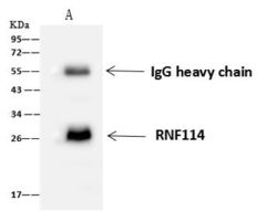
- Immunoprecipitation of ZNF313 was performed on (Lane A) 0.5 mg 293T whole cell lysate using 4 µL of ZNF313 Polyclonal Antibody (Product # PA5-112261) at a dilution of 1:100, and 60 µg of Immunomagnetic beads Protein A/G. A Goat Anti-Rabbit IgG (H+L)/HRP was used as a secondary antibody at a dilution of 1:10,000. Developed using the ECL technique and performed under reducing conditions. Predicted band size: 26 kDa. Observed band size: 26 kDa.
