 Explore
 Validate
 Learn
Learn Western blot
Antibody data
- Antibody Data
- Antigen structure
- References [0]
- Comments [0]
- Validations
- Western blot [3]
- Immunocytochemistry [2]
- Immunohistochemistry [2]
Submit
Validation data
Reference
Comment
Report error
- Product number
- PA5-22367 - Provider product page

- Provider
- Invitrogen Antibodies
- Product name
- HSD17B4 Polyclonal Antibody
- Antibody type
- Polyclonal
- Antigen
- Recombinant protein fragment
- Description
- Recommended positive controls: HeLa, mouse brain. Predicted reactivity: Mouse (92%), Rat (92%), Zebrafish (85%), Xenopus laevis (81%), Pig (87%), Chicken (84%), Bovine (87%), Guinea pig (85%). Store product as a concentrated solution. Centrifuge briefly prior to opening the vial.
- Reactivity
- Human, Mouse
- Host
- Rabbit
- Isotype
- IgG
- Vial size
- 100 µL
- Concentration
- 0.66 mg/mL
- Storage
- Store at 4°C short term. For long term storage, store at -20°C, avoiding freeze/thaw cycles.
No comments: Submit comment
Supportive validation
- Submitted by
- Invitrogen Antibodies (provider)
- Main image

- Experimental details
- Western Blot using HSD17B4 Polyclonal Antibody (Product # PA5-22367). Sample (50 µg of whole cell lysate). Lane A: Mouse brain . 7.5% SDS PAGE. HSD17B4 Polyclonal Antibody (Product # PA5-22367) diluted at 1:1,000.

- Submitted by
- Invitrogen Antibodies (provider)
- Main image

- Experimental details
- Western Blot using HSD17B4 Polyclonal Antibody (Product # PA5-22367). Sample (30 µg of whole cell lysate). Lane A: Hela . 7.5% SDS PAGE. HSD17B4 Polyclonal Antibody (Product # PA5-22367) diluted at 1:1,000.

- Submitted by
- Invitrogen Antibodies (provider)
- Main image

- Experimental details
- Western blot was performed using Anti-HSD17B4 Polyclonal Antibody (Product # PA5-22367) and an 80 kDa band corresponding to HSD17B4 was observed in all tissue lysates tested except Mouse Brain. Lower expression of SAMHD1 was observed in Mouse Brain as compared to Mouse Testis and Mouse Heart [10.1016/s0960-0760(99)00066-7]. Tissue lysates (30g lysate) of Mouse Testis (Lane 1), Mouse Heart (Lane 2) and Mouse Brain (Lane 3) were electrophoresed using Novex® NuPAGE® 4-12 % Bis-Tris gel (Product # NP0322BOX). Resolved proteins were then transferred onto a nitrocellulose membrane (Product # IB23001) by iBlot® 2 Dry Blotting System (Product # IB21001). The blot was probed with the primary antibody (1:2000 dilution) and detected by chemiluminescence with Goat anti-Rabbit IgG (H+L), Superclonal™ Recombinant Secondary Antibody, HRP (Product # A27036, 1:4000 dilution) using the iBright FL 1000 (Product # A32752). Chemiluminescent detection was performed using Novex® ECL Chemiluminescent Substrate Reagent Kit (Product # WP20005). Two uncharacterized bands (*) were observed in certain models.

Supportive validation
- Submitted by
- Invitrogen Antibodies (provider)
- Main image

- Experimental details
- Immunocytochemistry-Immunofluorescence analysis of HSD17B4 was performed in HeLa cells fixed in ice-cold MeOH for 5 min. Green: HSD17B4 Polyclonal Antibody (Product # PA5-22367) diluted at 1:500. Blue: Hoechst 33342 staining.

Supportive validation
- Submitted by
- Invitrogen Antibodies (provider)
- Main image

- Experimental details
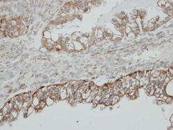
- Immunohistochemical analysis of paraffin-embedded human ovarian cancer, using HSD17B4 (Product # PA5-22367) antibody at 1:500 dilution. Antigen Retrieval: EDTA based buffer, pH 8.0, 15 min.

- Submitted by
- Invitrogen Antibodies (provider)
- Main image
- Experimental details
- Immunohistochemical analysis of paraffin-embedded human ovarian cancer, using HSD17B4 (Product # PA5-22367) antibody at 1:500 dilution. Antigen Retrieval: EDTA based buffer, pH 8.0, 15 min.
