 Explore
 Validate
 Learn
Learn Western blot
 Immunohistochemistry
Antibody data
- Antibody Data
- Antigen structure
- References [1]
- Comments [0]
- Validations
- Western blot [1]
- Immunohistochemistry [7]
Submit
Validation data
Reference
Comment
Report error
- Product number
- HPA004728 - Provider product page

- Provider
- Atlas Antibodies
- Proper citation
- Atlas Antibodies Cat#HPA004728, RRID:AB_1080360
- Product name
- Anti-TGM3
- Antibody type
- Polyclonal
- Reactivity
- Human
- Host
- Rabbit
- Conjugate
- Unconjugated
- Antigen sequence
GSDQERQVFQKALGKLKPNTPFAATSSMGLETEEQ
EPSIIGKLKVAGMLAVGKEVNLVLLLKNLSRDTKT
VTVNMTAWTIIYNGTLVHEVWKDSATMSLDPEEEA
EHPIKISYAQYEKYLKSDNMI- Isotype
- IgG
- Vial size
- 100 µl
- Storage
- Store at +4°C for short term storage. Long time storage is recommended at -20°C.
Submitted references Transglutaminase 3 as a prognostic biomarker in esophageal cancer revealed by proteomics
Uemura N, Nakanishi Y, Kato H, Saito S, Nagino M, Hirohashi S, Kondo T
International Journal of Cancer 2009 May;124(9):2106-2115
International Journal of Cancer 2009 May;124(9):2106-2115
No comments: Submit comment
Supportive validation
- Submitted by
- Atlas Antibodies (provider)
- Main image

- Experimental details
- Western blot analysis in human cell line RT-4, human cell line U-251 MG, human plasma, human liver tissue and human tonsil tissue.

Enhanced validation
Supportive validation
- Submitted by
- Atlas Antibodies (provider)
- Enhanced method
- Orthogonal validation
- Main image

- Experimental details
- Immunohistochemistry analysis in human esophagus and stomach tissues using HPA004728 antibody. Corresponding TGM3 RNA-seq data are presented for the same tissues.
- Sample type
- HUMAN

Supportive validation
- Submitted by
- Atlas Antibodies (provider)
- Main image

- Experimental details
- Immunohistochemical staining of human esophagus shows high expression.
- Sample type
- HUMAN
- Submitted by
- Atlas Antibodies (provider)
- Main image

- Experimental details
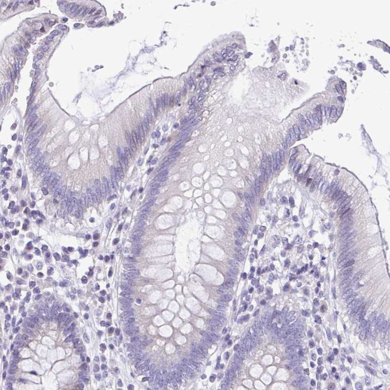
- Immunohistochemical staining of human colon shows low expression as expected.
- Sample type
- HUMAN
- Submitted by
- Atlas Antibodies (provider)
- Main image

- Experimental details
- Immunohistochemical staining of human lymph node shows no positivity in germinal center cells and non-germinal center cells as expected.
- Sample type
- HUMAN

- Submitted by
- Atlas Antibodies (provider)
- Main image
- Experimental details
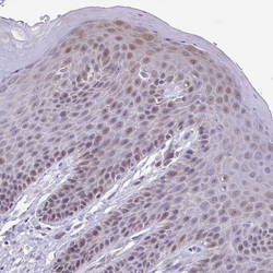
- Immunohistochemical staining of human skin shows weak cytoplasmic positivity in squamous epithelial cells.
- Sample type
- HUMAN

- Submitted by
- Atlas Antibodies (provider)
- Main image

- Experimental details
- Immunohistochemical staining of human stomach shows no positivity in glandular cells as expected.
- Sample type
- HUMAN

- Submitted by
- Atlas Antibodies (provider)
- Main image

- Experimental details
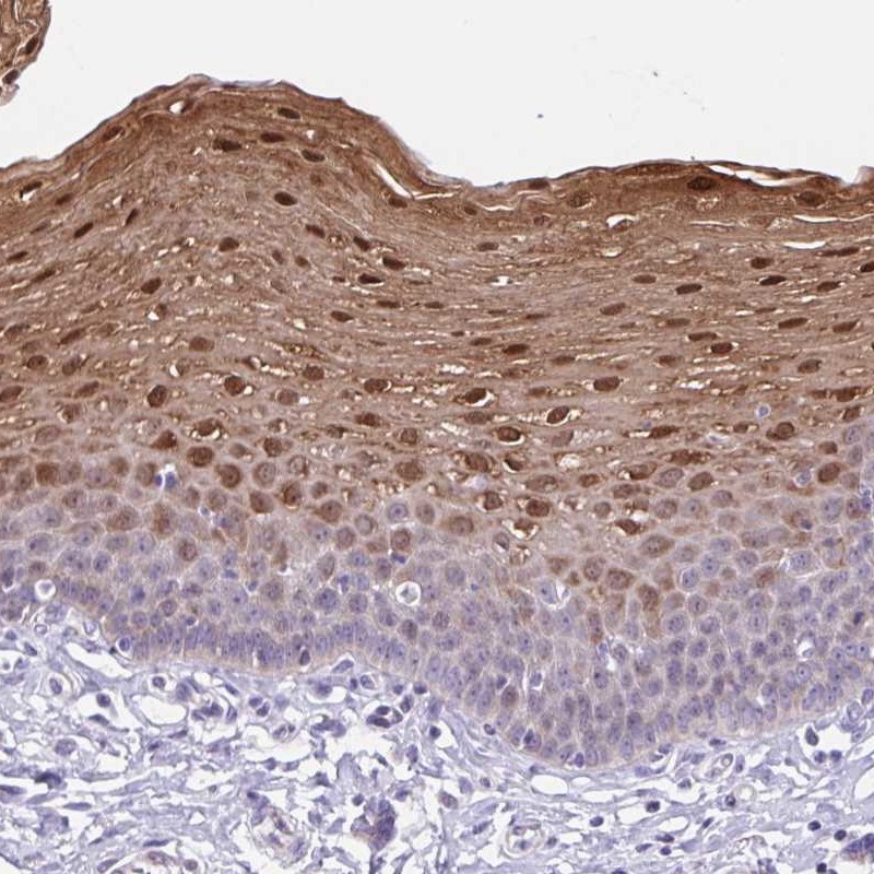
- Immunohistochemical staining of human esophagus shows strong cytoplasmic and nuclear positivity in squamous epithelial cells.
- Sample type
- HUMAN
