Antibody data
- Antibody Data
- Antigen structure
- References [1]
- Comments [0]
- Validations
- Immunocytochemistry [1]
- Immunohistochemistry [1]
- Other assay [1]
Submit
Validation data
Reference
Comment
Report error
- Product number
- PA5-97527 - Provider product page

- Provider
- Invitrogen Antibodies
- Product name
- Carbonic Anhydrase I Polyclonal Antibody
- Antibody type
- Polyclonal
- Antigen
- Recombinant full-length protein
- Reactivity
- Human, Mouse
- Host
- Rabbit
- Isotype
- IgG
- Vial size
- 100 µg
- Concentration
- 3.3 mg/mL
- Storage
- -20°C or -80°C if preferred
Submitted references Single-cell atlas of the aging mouse colon.
Širvinskas D, Omrani O, Lu J, Rasa M, Krepelova A, Adam L, Kaeppel S, Sommer F, Neri F
iScience 2022 May 20;25(5):104202
iScience 2022 May 20;25(5):104202
No comments: Submit comment
Supportive validation
- Submitted by
- Invitrogen Antibodies (provider)
- Main image

- Experimental details
- Immunofluorescent analysis of Carbonic Anhydrase I in A549 cells using a Carbonic Anhydrase I polyclonal antibody (Product # PA5-97527) at a dilution of 1:100. Alexa Fluor 488-congugated Goat Anti-Rabbit IgG(H+L) secondary antibody was used.
Supportive validation
- Submitted by
- Invitrogen Antibodies (provider)
- Main image

- Experimental details
- Immunohistochemical analysis of Carbonic Anhydrase I in paraffin embedded human melanoma using a Carbonic Anhydrase I polyclonal antibody (Product # PA5-97527) at a dilution of 1:100.
Supportive validation
- Submitted by
- Invitrogen Antibodies (provider)
- Main image

- Experimental details
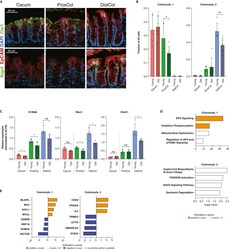
- Changes in colonocytes during aging (A) Immunofluorescent staining of different compartments for Car1 (upper) and Aqp4 (lower). EpCAM staining used to stain epithelial cells, DAPI used to stain nuclei. Scale bar 50 mum. (B) Bar charts of colonocyte_1 and _2 fractions in different compartments in young and old. (n = 5 young, six old biologically independent animals, 1-tailed unpaired t -test used between young and old animals; Data are represented as mean +SD). (C) qPCR of colonocyte-specific genes between young and old female mice. Young samples are taken from Figure S1 to be used as controls. (n = 4 young, 3 old biologically independent animals; 1-tailed unpaired t -test used between young and old animals; Data are represented as mean +SD). (D) Enriched Canonical Pathways from IPA for colonocytes (n = 5 young, 6 old biologically independent animals). (E) Predicted Upstream Regulators from IPA for colonocytes (n = 5 young, 6 old biologically independent animals). ns, p>=0.05; *, p < 0.05; **, p < 0.01; ***, p < 0.001.
 Explore
 Validate
 Learn
Learn Western blot
 ELISA
ELISA Immunocytochemistry