 Explore
 Validate
 Learn
LearnGTX12412
antibody from GeneTex
Targeting: PPARA
hPPAR, NR1C1, PPAR
 Western blot
 Immunocytochemistry Immunoprecipitation Flow cytometry Gel shift Chromatin Immunoprecipitation
Antibody data
- Antibody Data
- Antigen structure
- References [0]
- Comments [0]
- Validations
- Western blot [2]
- Flow cytometry [4]
Submit
Validation data
Reference
Comment
Report error
- Product number
- GTX12412 - Provider product page

- Provider
- GeneTex
- Proper citation
- GeneTex Cat#GTX12412, RRID:AB_371701
- Product name
- PPAR alpha antibody [3B6/PPAR]
- Antibody type
- Monoclonal
- Reactivity
- Human, Mouse, Rat, Bovine, Rabbit
- Host
- Mouse
No comments: Submit comment
Supportive validation
- Submitted by
- GeneTex (provider)
- Main image

- Experimental details
- Western blot analysis of PPAR alpha in 25 ug of HeLa, Jurkat and NIH-3T3 cell lysates. Proteins were transferred to a PVDF membrane and blocked with a blocking buffer at 4oC overnight. The membrane was probed with PPAR alpha antibody [3B6/PPAR] at a dilution of 1:1000 (HeLa and Jurkat) and 1:500 overnight at 4¢XC, washed in TBST, and probed with a proper secondary antibody for 1 hr at room temperature in the dark. Chemiluminescent detection was performed.

- Submitted by
- GeneTex (provider)
- Main image

- Experimental details
- Western blot analysis of PPAR alpha was performed by loading 25 ug of Hela (Lane 1), Jurkat (Lane 2), and NIH-3T3 cell lysates (Lane 3) and a molecular weight protein ladder onto an SDS polyacrylamide gel. Proteins were transferred to a PVDF membrane and blocked with a blocking buffer at 4ºC overnight. The membrane was probed with a PPAR alpha monoclonal antibody (GTX12412) at a dilution of 1:1000 (Hela and Jurkat) and 1:500 (NIH-3T3) overnight at 4°C, washed in TBST, and probed with an HRP-conjugated secondary antibody for 1 hr at room temperature in the dark. Chemiluminescent detection was performed using ECL substrate. Results show a band at 52 kDa in all three cell lines.
- Validation comment
- WB

Supportive validation
- Submitted by
- GeneTex (provider)
- Main image
- Experimental details
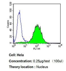
- Flow cytometry analysis of PPAR alpha in Hela cells compared to an isotype control (blue). Cells were harvested, adjusted to a concentration of 1-5x10^6 cells/ml, fixed with 2% paraformaldehyde, washed with PBS, and incubated with PPAR alpha antibody [3B6/PPAR] at a dilution of 0.25 ug/test for 60 min at room temperature. Cells were then blocked in a solution of 2% BSA-PBS for 30 min at room temperature, incubated for 40 min at room temperature in the dark with a proper secondary antibody, and re-suspended in PBS for FACS analysis.

- Submitted by
- GeneTex (provider)
- Main image
- Experimental details
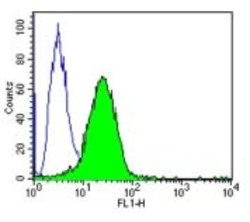
- Flow cytometry analysis of PPAR alpha showing positive staining in the nucleus of 3T3 cells compared to an isotype control (blue). Cells were harvested, adjusted to a concentration of 1-5x10^6 cells/ml, fixed with 2% paraformaldehyde, washed with PBS, and incubated with PPAR alpha monoclonal antibody (GTX12412) at a dilution of 0.25 ug/test for 60 min at room temperature. Cells were then blocked in a solution of 2% BSA-PBS for 30 min at room temperature, incubated for 40 min at room temperature in the dark using a Dylight 488-conjugated goat anti-mouse IgG (H+L) secondary antibody, and re-suspended in PBS for FACS analysis.
- Validation comment
- FACS

- Submitted by
- GeneTex (provider)
- Main image
- Experimental details
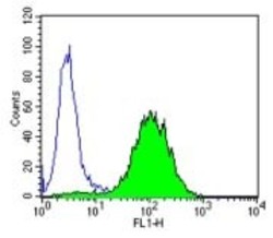
- Flow cytometry analysis of PPAR alpha showing positive staining in the nucleus of Hela cells compared to an isotype control (blue). Cells were harvested, adjusted to a concentration of 1-5x10^6 cells/ml, fixed with 2% paraformaldehyde, washed with PBS, and incubated with PPAR alpha monoclonal antibody (GTX12412) at a dilution of 0.25 ug/test for 60 min at room temperature. Cells were then blocked in a solution of 2% BSA-PBS for 30 min at room temperature, incubated for 40 min at room temperature in the dark using a Dylight 488-conjugated goat anti-mouse IgG (H+L) secondary antibody, and re-suspended in PBS for FACS analysis.
- Validation comment
- FACS

- Submitted by
- GeneTex (provider)
- Main image

- Experimental details
- Flow cytometry analysis of PPAR alpha showing positive staining in the nucleus of Jurkat cells compared to an isotype control (blue). Cells were harvested, adjusted to a concentration of 1-5x10^6 cells/ml, fixed with 2% paraformaldehyde, washed with PBS, and incubated with PPAR alpha monoclonal antibody (GTX12412) at a dilution of 0.25 ug/test for 60 min at room temperature. Cells were then blocked in a solution of 2% BSA-PBS for 30 min at room temperature, incubated for 40 min at room temperature in the dark using a Dylight 488-conjugated goat anti-mouse IgG (H+L) secondary antibody, and re-suspended in PBS for FACS analysis.
- Validation comment
- FACS
