Explore
 Validate
 Learn
Learn Immunocytochemistry
 Immunohistochemistry
Antibody data
- Antibody Data
- Antigen structure
- References [1]
- Comments [0]
- Validations
- Immunocytochemistry [1]
- Immunohistochemistry [7]
Submit
Validation data
Reference
Comment
Report error
- Product number
- HPA020646 - Provider product page

- Provider
- Atlas Antibodies
- Proper citation
- Atlas Antibodies Cat#HPA020646, RRID:AB_1856752
- Product name
- Anti-SETD1A
- Antibody type
- Polyclonal
- Reactivity
- Human
- Host
- Rabbit
- Conjugate
- Unconjugated
- Antigen sequence
AFGAFDQWWESKEEKAKPFQNAAKQQAKEEDKEKT
KLKEPGLLSLVDWAKSGGTTGIEAFAFGSGLRGAL
RLPSFKVKRKEPSEISEASEEKRPRPSTPAEEDED
DP- Isotype
- IgG
- Vial size
- 100 µl
- Storage
- Store at +4°C for short term storage. Long time storage is recommended at -20°C.
Submitted references Enhanced HSP70 lysine methylation promotes proliferation of cancer cells through activation of Aurora kinase B.
Cho HS, Shimazu T, Toyokawa G, Daigo Y, Maehara Y, Hayami S, Ito A, Masuda K, Ikawa N, Field HI, Tsuchiya E, Ohnuma S, Ponder BA, Yoshida M, Nakamura Y, Hamamoto R
Nature communications 2012;3:1072
Nature communications 2012;3:1072
No comments: Submit comment
Supportive validation
- Submitted by
- Atlas Antibodies (provider)
- Main image

- Experimental details
- Immunofluorescent staining of human cell line A-431 shows localization to nuclear speckles.
- Sample type
- HUMAN

Enhanced validation
Enhanced validation
Supportive validation
- Submitted by
- Atlas Antibodies (provider)
- Enhanced method
- Orthogonal validation
- Main image
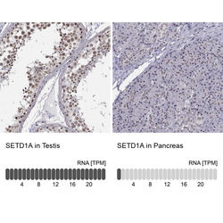
- Experimental details
- Immunohistochemistry analysis in human testis and pancreas tissues using Anti-SETD1A antibody. Corresponding SETD1A RNA-seq data are presented for the same tissues.
- Sample type
- HUMAN

Enhanced validation
- Submitted by
- Atlas Antibodies (provider)
- Enhanced method
- Independent antibody validation
- Main image

- Experimental details
- Immunohistochemical staining of human cerebral cortex, kidney, pancreas and testis using Anti-SETD1A antibody HPA020646 (A) shows similar protein distribution across tissues to independent antibody HPA058376 (B).

Supportive validation
- Submitted by
- Atlas Antibodies (provider)
- Main image

- Experimental details
- Immunohistochemical staining of human small intestine shows nuclear positivity in glandular cells.

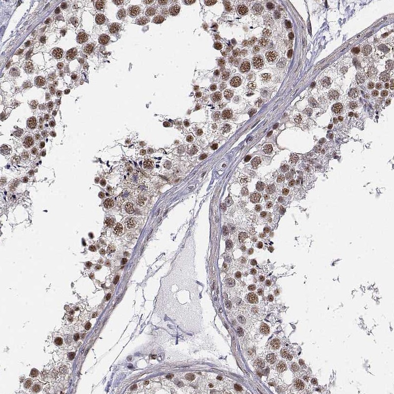
- Submitted by
- Atlas Antibodies (provider)
- Main image

- Experimental details
- Immunohistochemical staining of human testis shows high expression.
- Sample type
- HUMAN
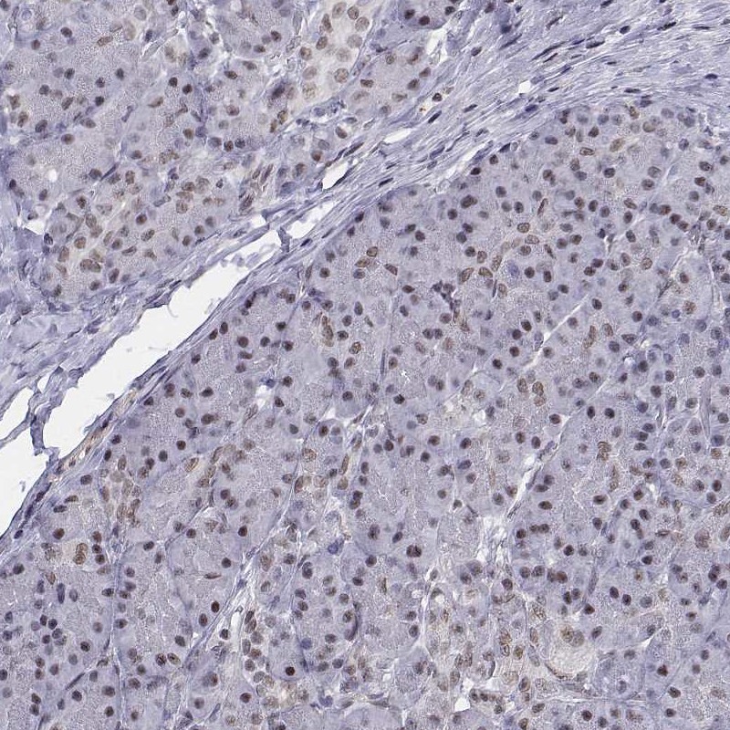
- Submitted by
- Atlas Antibodies (provider)
- Main image
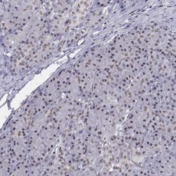
- Experimental details
- Immunohistochemical staining of human pancreas shows low expression as expected.
- Sample type
- HUMAN
- Submitted by
- Atlas Antibodies (provider)
- Main image

- Experimental details
- Immunohistochemical staining of human kidney using Anti-SETD1A antibody HPA020646.
- Sample type
- HUMAN

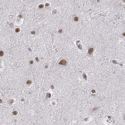
- Submitted by
- Atlas Antibodies (provider)
- Main image

- Experimental details
- Immunohistochemical staining of human cerebral cortex using Anti-SETD1A antibody HPA020646.
- Sample type
- HUMAN