 Explore
 Validate
 Learn
Learn Western blot
Antibody data
- Antibody Data
- Antigen structure
- References [5]
- Comments [0]
- Validations
- Western blot [3]
- Immunoprecipitation [1]
- Immunohistochemistry [3]
- Chromatin Immunoprecipitation [1]
Submit
Validation data
Reference
Comment
Report error
- Product number
- NB100-558 - Provider product page

- Provider
- Novus Biologicals
- Proper citation
- Novus Cat#NB100-558, RRID:AB_2185760
- Product name
- Rabbit Polyclonal SETD1A Antibody
- Antibody type
- Polyclonal
- Description
- Immunogen affinity purified.
- Reactivity
- Human
- Host
- Rabbit
- Isotype
- IgG
- Vial size
- 100 ul
- Concentration
- 1.0 mg/ml
- Storage
- Store at 4C. Do not freeze.
Submitted references SIRT1 deacetylase promotes acquisition of genetic mutations for drug resistance in CML cells.
Contribution of H3K4 methylation by SET-1A to interleukin-1-induced cyclooxygenase 2 and inducible nitric oxide synthase expression in human osteoarthritis chondrocytes.
Impaired recruitment of the histone methyltransferase DOT1L contributes to the incomplete reactivation of tumor suppressor genes upon DNA demethylation.
Transient high glucose causes persistent epigenetic changes and altered gene expression during subsequent normoglycemia.
Identification and characterization of the human Set1B histone H3-Lys4 methyltransferase complex.
Wang Z, Yuan H, Roth M, Stark JM, Bhatia R, Chen WY
Oncogene 2013 Jan 31;32(5):589-98
Oncogene 2013 Jan 31;32(5):589-98
Contribution of H3K4 methylation by SET-1A to interleukin-1-induced cyclooxygenase 2 and inducible nitric oxide synthase expression in human osteoarthritis chondrocytes.
El Mansouri FE, Chabane N, Zayed N, Kapoor M, Benderdour M, Martel-Pelletier J, Pelletier JP, Duval N, Fahmi H
Arthritis and rheumatism 2011 Jan;63(1):168-79
Arthritis and rheumatism 2011 Jan;63(1):168-79
Impaired recruitment of the histone methyltransferase DOT1L contributes to the incomplete reactivation of tumor suppressor genes upon DNA demethylation.
Jacinto FV, Ballestar E, Esteller M
Oncogene 2009 Nov 26;28(47):4212-24
Oncogene 2009 Nov 26;28(47):4212-24
Transient high glucose causes persistent epigenetic changes and altered gene expression during subsequent normoglycemia.
El-Osta A, Brasacchio D, Yao D, Pocai A, Jones PL, Roeder RG, Cooper ME, Brownlee M
The Journal of experimental medicine 2008 Sep 29;205(10):2409-17
The Journal of experimental medicine 2008 Sep 29;205(10):2409-17
Identification and characterization of the human Set1B histone H3-Lys4 methyltransferase complex.
Lee JH, Tate CM, You JS, Skalnik DG
The Journal of biological chemistry 2007 May 4;282(18):13419-28
The Journal of biological chemistry 2007 May 4;282(18):13419-28
No comments: Submit comment
Supportive validation
- Submitted by
- Novus Biologicals (provider)
- Main image
- Experimental details
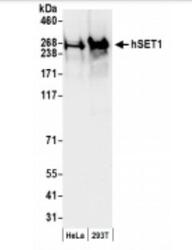
- Western Blot: SETD1A Antibody [NB100-558] - Nuclear extract (50 ug) from HeLa and 293T cells. Antibody: Affinity purified rabbit anti-hSET1 antibody used for WB at 0.1 ug/ml. Detection: Chemiluminescence with an exposure time of 10 seconds.

- Submitted by
- Novus Biologicals (provider)
- Main image

- Experimental details
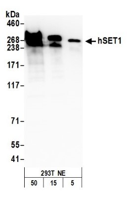
- Western Blot: SETD1A Antibody [NB100-558] - Detection of Human hSET1 by Western Blot. Samples: Nuclear extract (50, 15, 5 ug) from 293T cells. Antibody: Affinity purified rabbit anti-hSET1 antibody NB100-558 used for WB at 0.1 ug/ml. Detection: Chemiluminescence with an exposure time of 3 minutes.
- Submitted by
- Novus Biologicals (provider)
- Main image
- Experimental details
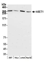
- Western Blot: SETD1A Antibody [NB100-558] - Detection of human hSET1 by western blot. Samples: Whole cell lysate (10 ug) from HEK293T, HeLa, Jurkat, and Hep-G2 cells prepared using NETN lysis buffer. Antibody: Affinity purified rabbit anti-hSET1 antibody A300-289A (lot A300-289A-6) used for WB at 0.04 ug/ml. Detection: Chemiluminescence with an exposure time of 75 seconds.

Supportive validation
- Submitted by
- Novus Biologicals (provider)
- Main image

- Experimental details
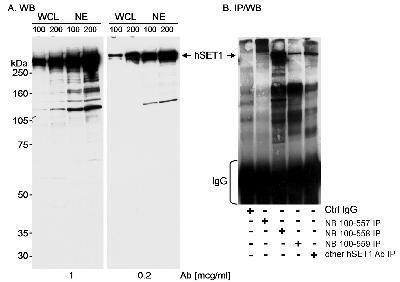
- Immunoprecipitation: SETD1A Antibody [NB100-558] - Detection of Human hSET1 on HeLa whole cell lysate using NB100-558. Affinity purified anti-hSET1 antibodies NB100-557, NB100-558, NB100-559, and another hSET1 antibody.
Supportive validation
- Submitted by
- Novus Biologicals (provider)
- Main image

- Experimental details
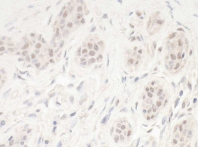
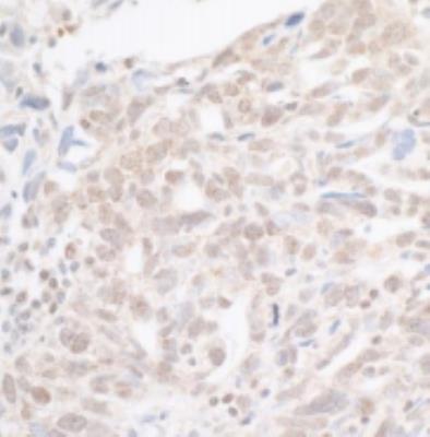
- Immunohistochemistry: SETD1A Antibody [NB100-558] - Sample: FFPE section of human ovarian carcinoma.~ Antibody: Affinity purified rabbit anti-hSet1 ~Cat. No. Lot4 used at a dilution of 1:1,000 (1ug/ml). Detection: DAB. Counterstain: IHC Hematoxylin (blue).

- Submitted by
- Novus Biologicals (provider)
- Main image

- Experimental details
- Immunohistochemistry: SETD1A Antibody [NB100-558] - Detection of human hSET by immunohistochemistry. Sample: FFPE section of human breast carcinoma. Antibody: Affinity purified rabbit anti-hSet1 NB100-558 used at 1:5,000 (0.2 µg/ml). Secondary: HRP-conjugated goat anti-rabbit IgG. Substrate: DAB.
- Submitted by
- Novus Biologicals (provider)
- Main image

- Experimental details
- Immunohistochemistry-Paraffin: SETD1A Antibody [NB100-558] - Section of human ovarian carcinoma. Antibody: Affinity purified rabbit anti-hSET1 used at 1:5,000 (0.2 ug/ml). Secondary: HRP-conjugated goat anti-rabbit IgG. Substrate: DAB.
Supportive validation
- Submitted by
- Novus Biologicals (provider)
- Main image

- Experimental details
- Chromatin Immunoprecipitation: SETD1A Antibody [NB100-558] - Chromatin Immunoprecipitation: SETD1A Antibody [NB100-558] - ChIP-chip scatter plot of anti-hSET1 (NB100-558) enriched DNA binding sites versus input reference DNA. A. 10 mcg of NB100-558 was used to immunoprecipitate chromatin from K562 cells per Ren et al (Genes Dev. 2002 16: 245-256). Immunoprecipitated DNA and reference DNA were amplified via ligation-mediated PCR and the products labeled with fluorescent dUTPs. The labeled ChIP and reference DNA were pooled, hybridized to a DNA microarray, and analyzed. Data points below the +3 SD curve (red line) represent significantly enriched binding sites. B. As a control, a similar experiment was performed using normal rabbit IgG. Compared to the anti-hSET1 ChIP, normal rabbit IgG showed little enrichment.
