 Explore
 Validate
 Learn
Learn Western blot
 Immunocytochemistry
Antibody data
- Antibody Data
- Antigen structure
- References [0]
- Comments [0]
- Validations
- Immunocytochemistry [6]
- Immunohistochemistry [1]
Submit
Validation data
Reference
Comment
Report error
- Product number
- 720030 - Provider product page

- Provider
- Invitrogen Antibodies
- Product name
- SALL4 Polyclonal Antibody
- Antibody type
- Polyclonal
- Antigen
- Synthetic peptide
- Description
- This antibody is predicted to react with Monkey, Rat, Mouse, Pig, Bovine, Rabbit and Dog. 720030 can be used for immunofluorescence analysis of Sall4 in human iPSCs and human ESC lines.
- Reactivity
- Human, Mouse
- Host
- Rabbit
- Isotype
- IgG
- Vial size
- 100 μg
- Concentration
- 0.5 mg/mL
- Storage
- Store at 4°C short term. For long term storage, store at -20°C, avoiding freeze/thaw cycles.
No comments: Submit comment
Supportive validation
- Submitted by
- Invitrogen Antibodies (provider)
- Main image
- Experimental details
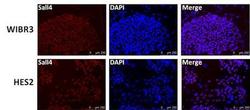
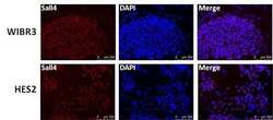
- Immunofluorescent analysis of Sall4 (red) in human ESCs WIRB3 and HES2. The cells were fixed with 300 uL 4% paraformaldehyde solution for 15 min at RT in the dark, permeabilized with 300 uL 0.25% TritonX-100 solution (diluted in PBS) for 5 min at RT, and blocked with 300 uL 10% BSA solution (diluted in PBS) for 30 min in a 37C incubator. Cells were stained with a Sall4 polyclonal antibody (Product # 720030) at a dilution of 1:250 (2 µg/mL) in 3% BSA solution (diluted in PBS) at 4C overnight, and then incubated with a secondary antibody (Red). Nuclei (blue) was stained with DAPI (Product # D1306). Images were taken on a Leica DM5500 microscope at 10X magnification. Data courtesy of Dr. Jianlong Wang's lab.

- Submitted by
- Invitrogen Antibodies (provider)
- Main image
- Experimental details
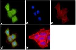
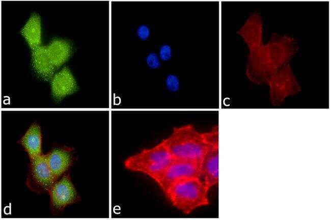
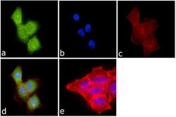
- Immunofluorescence was performed on fixed and permeabilized A-431cells for detection of Sall4 using Anti-Sall4 Rabbit Polyclonal Antibody (Product # 720030, 2 µg/mL) and labeled with Goat anti-Rabbit IgG (H+L) Superclonal Secondary Antibody, Alexa Fluor® 488 conjugate (Product # A27034, 0.4 µg/mL, 1:2500). Panel a) shows representative cells that were stained for detection and localization of Sall4 protein (green), Panel b) is stained for nuclei (blue) using SlowFade® Gold Antifade Mountant with DAPI (Product # S36938, 1:50). Panel c) represents cytoskeletal F-actin staining using Alexa Fluor® 594 Phalloidin (Product # A12381, 1:200). Panel d) is a composite image of Panels a, b and c clearly demonstrating nuclear localization of Sall4. Panel e) represents control cells with no primary antibody to assess background.
- Submitted by
- Invitrogen Antibodies (provider)
- Main image

- Experimental details
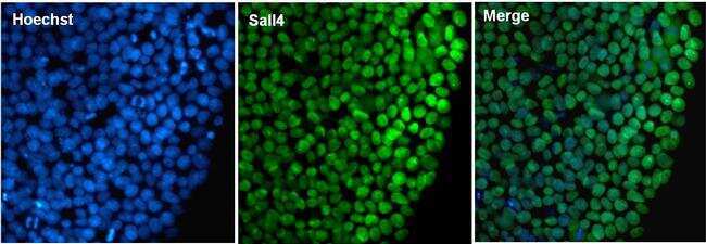
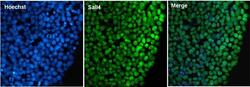
- Immunofluorescent analysis of Sall4 (green) in Episomal Human iPSC cells. The cells were fixed with formalin for 15 minutes, permeabilized with 0.1% Triton X-100 in PBS for 15 minutes, and blocked with 3% BSA in PBS (Product # 37525) for 30 minutes at room temperature. Cells were stained with a Sall4 polyclonal antibody (Product # 720030), at a dilution of 2 µg/mL for one hour at room temperature, and then incubated with a goat anti-rabbit Superclonal IgG secondary antibody, Alexa Fluor 488 conjugate (Product # A27034) at a dilution of 1:1000 for 30 minutes at room temperature (green). Nuclei (blue) were counterstained with Hoechst 33342 dye (Product # 62249). Images were taken on a Thermo Scientific EVOS FL at 40X magnification.
- Submitted by
- Invitrogen Antibodies (provider)
- Main image
- Experimental details
- Immunofluorescent analysis of Sall4 (red) in human ESCs WIRB3 and HES2. The cells were fixed with 300 uL 4% paraformaldehyde solution for 15 min at RT in the dark, permeabilized with 300 uL 0.25% TritonX-100 solution (diluted in PBS) for 5 min at RT, and blocked with 300 uL 10% BSA solution (diluted in PBS) for 30 min in a 37C incubator. Cells were stained with a Sall4 polyclonal antibody (Product # 720030) at a dilution of 1:250 (2 µg/mL) in 3% BSA solution (diluted in PBS) at 4C overnight, and then incubated with a secondary antibody (Red). Nuclei (blue) was stained with DAPI (Product # D1306). Images were taken on a Leica DM5500 microscope at 10X magnification. Data courtesy of Dr. Jianlong Wang's lab.

- Submitted by
- Invitrogen Antibodies (provider)
- Main image
- Experimental details
- Immunofluorescent analysis of Sall4 (green) in Episomal Human iPSC cells. The cells were fixed with formalin for 15 minutes, permeabilized with 0.1% Triton X-100 in PBS for 15 minutes, and blocked with 3% BSA in PBS (Product # 37525) for 30 minutes at room temperature. Cells were stained with a Sall4 polyclonal antibody (Product # 720030), at a dilution of 2 µg/mL for one hour at room temperature, and then incubated with a goat anti-rabbit Superclonal IgG secondary antibody, Alexa Fluor 488 conjugate (Product # A27034) at a dilution of 1:1000 for 30 minutes at room temperature (green). Nuclei (blue) were counterstained with Hoechst 33342 dye (Product # 62249). Images were taken on a Thermo Scientific EVOS FL at 40X magnification.

- Submitted by
- Invitrogen Antibodies (provider)
- Main image
- Experimental details
- Immunofluorescence was performed on fixed and permeabilized A-431cells for detection of Sall4 using Anti-Sall4 Rabbit Polyclonal Antibody (Product # 720030, 2 µg/mL) and labeled with Goat anti-Rabbit IgG (Heavy Chain) Superclonal Secondary Antibody, Alexa Fluor® 488 conjugate (Product # A27034, 0.4 µg/mL, 1:2500). Panel a) shows representative cells that were stained for detection and localization of Sall4 protein (green), Panel b) is stained for nuclei (blue) using SlowFade® Gold Antifade Mountant with DAPI (Product # S36938, 1:50). Panel c) represents cytoskeletal F-actin staining using Alexa Fluor® 594 Phalloidin (Product # A12381, 1:200). Panel d) is a composite image of Panels a, b and c clearly demonstrating nuclear localization of Sall4. Panel e) represents control cells with no primary antibody to assess background.

Supportive validation
- Submitted by
- Invitrogen Antibodies (provider)
- Main image
- Experimental details
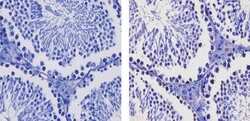
- Immunohistochemistry analysis of Sall4 showing staining in the nucleus of paraffin-embedded mouse testis tissue (right) compared to a negative control without primary antibody (left). To expose target proteins, antigen retrieval was performed using 10mM sodium citrate (pH 6.0), microwaved for 8-15 min. Following antigen retrieval, tissues were blocked in 3% H2O2-methanol for 15 min at room temperature, washed with ddH2O and PBS, and then probed with a Sall4 Rabbit Polyclonal Antibody (Product # 720030) diluted in 3% BSA-PBS at a dilution of 1:100 for 1 hour at 37ºC in a humidified chamber. Tissues were washed extensively in PBST and detection was performed using an HRP-conjugated secondary antibody followed by colorimetric detection using a DAB kit. Tissues were counterstained with hematoxylin and dehydrated with ethanol and xylene to prep for mounting.
