Antibody data
- Antibody Data
- Antigen structure
- References [6]
- Comments [0]
- Validations
- Western blot [5]
- Immunohistochemistry [4]
- Other assay [6]
Submit
Validation data
Reference
Comment
Report error
- Product number
- PA5-29559 - Provider product page

- Provider
- Invitrogen Antibodies
- Product name
- ANP Polyclonal Antibody
- Antibody type
- Polyclonal
- Antigen
- Recombinant full-length protein
- Description
- Recommended positive controls: mouse heart, rat heart. Predicted reactivity: Mouse (80%), Rat (80%), Dog (85%), Pig (87%), Rabbit (81%), Bovine (85%). Store product as a concentrated solution. Centrifuge briefly prior to opening the vial.
- Reactivity
- Human, Mouse, Rat
- Host
- Rabbit
- Isotype
- IgG
- Vial size
- 100 µL
- Concentration
- 1.43 mg/mL
- Storage
- Store at 4°C short term. For long term storage, store at -20°C, avoiding freeze/thaw cycles.
Submitted references RIP3 Contributes to Cardiac Hypertrophy by Influencing MLKL-Mediated Calcium Influx.
Atrial Natriuretic Peptide Promotes Neurite Outgrowth and Survival of Cochlear Spiral Ganglion Neurons in vitro Through NPR-A/cGMP/PKG Signaling.
Vaspin Mediates the Intraorgan Crosstalk Between Heart and Adipose Tissue in Lipoatrophic Mice.
Natriuretic Peptides Regulate Prostate Cells Inflammatory Behavior: Potential Novel Anticancer Agents for Prostate Cancer.
Atrial Natriuretic Peptide Improves Neurite Outgrowth from Spiral Ganglion Neurons In Vitro through a cGMP-Dependent Manner.
Cardiac specific PRMT1 ablation causes heart failure through CaMKII dysregulation.
Xue H, Shi H, Zhang F, Li H, Li C, Han Q
Oxidative medicine and cellular longevity 2022;2022:5490553
Oxidative medicine and cellular longevity 2022;2022:5490553
Atrial Natriuretic Peptide Promotes Neurite Outgrowth and Survival of Cochlear Spiral Ganglion Neurons in vitro Through NPR-A/cGMP/PKG Signaling.
Sun F, Zhou K, Tian KY, Zhang XY, Liu W, Wang J, Zhong CP, Qiu JH, Zha DJ
Frontiers in cell and developmental biology 2021;9:681421
Frontiers in cell and developmental biology 2021;9:681421
Vaspin Mediates the Intraorgan Crosstalk Between Heart and Adipose Tissue in Lipoatrophic Mice.
Zhang D, Zhu H, Zhan E, Wang F, Liu Y, Xu W, Liu X, Liu J, Li S, Pan Y, Wang Y, Cao W
Frontiers in cell and developmental biology 2021;9:647131
Frontiers in cell and developmental biology 2021;9:647131
Natriuretic Peptides Regulate Prostate Cells Inflammatory Behavior: Potential Novel Anticancer Agents for Prostate Cancer.
Mezzasoma L, Talesa VN, Costanzi E, Bellezza I
Biomolecules 2021 May 25;11(6)
Biomolecules 2021 May 25;11(6)
Atrial Natriuretic Peptide Improves Neurite Outgrowth from Spiral Ganglion Neurons In Vitro through a cGMP-Dependent Manner.
Sun F, Zhou K, Tian KY, Wang J, Qiu JH, Zha DJ
Neural plasticity 2020;2020:8831735
Neural plasticity 2020;2020:8831735
Cardiac specific PRMT1 ablation causes heart failure through CaMKII dysregulation.
Pyun JH, Kim HJ, Jeong MH, Ahn BY, Vuong TA, Lee DI, Choi S, Koo SH, Cho H, Kang JS
Nature communications 2018 Nov 30;9(1):5107
Nature communications 2018 Nov 30;9(1):5107
No comments: Submit comment
Supportive validation
- Submitted by
- Invitrogen Antibodies (provider)
- Main image
- Experimental details
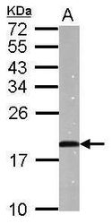
- Western blot analysis of ANP using 50 µg of mouse heart lysate. Samples were loaded onto a 15% SDS-PAGE gel and probed with an ANP polyclonal antibody (Product # PA5-29559) at a dilution of 1:5000.
- Submitted by
- Invitrogen Antibodies (provider)
- Main image

- Experimental details
- Western blot analysis of ANP using Mouse tissue extracts (50 µg). Samples were loaded onto a 15% SDS-PAGE gel and probed with an ANP polyclonal antibody (Product # PA5-29559) at a dilution of 1:5000.
- Submitted by
- Invitrogen Antibodies (provider)
- Main image

- Experimental details
- Western blot analysis of ANP was performed by separating 50 µg of mouse tissue extract by 15% SDS-PAGE. Proteins were transferred to a membrane and probed with a ANP Polyclonal Antibody (Product # PA5-29559) at a dilution of 1:20000. The HRP-conjugated anti-rabbit IgG antibody was used to detect the primary antibody.
- Submitted by
- Invitrogen Antibodies (provider)
- Main image

- Experimental details
- Western Blot using ANP Polyclonal Antibody (Product # PA5-29559). Various tissue extracts (50 µg) were separated by 15% SDS-PAGE, and the membrane was blotted with ANP Polyclonal Antibody (Product # PA5-29559) diluted at 1:2,000. The HRP-conjugated anti-rabbit IgG antibody was used to detect the primary antibody.
- Submitted by
- Invitrogen Antibodies (provider)
- Main image

- Experimental details
- Western blot analysis of ANP was performed by separating 50 µg of rat tissue extract by 15% SDS-PAGE. Proteins were transferred to a membrane and probed with a ANP Polyclonal Antibody (Product # PA5-29559) at a dilution of 1:2000. The HRP-conjugated anti-rabbit IgG antibody was used to detect the primary antibody.
Supportive validation
- Submitted by
- Invitrogen Antibodies (provider)
- Main image
- Experimental details
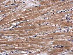
- Immunohistochemistry (Paraffin) analysis of ANP was performed in paraffin-embedded mouse muscle tissue using ANP Polyclonal Antibody (Product # PA5-29559) at a dilution of 1:500.
- Submitted by
- Invitrogen Antibodies (provider)
- Main image
- Experimental details
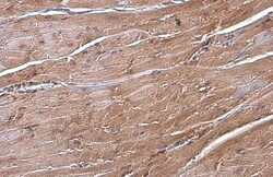
- Immunohistochemistry (Paraffin) analysis of ANP was performed in paraffin-embedded mouse heart tissue using ANP Polyclonal Antibody (Product # PA5-29559) at a dilution of 1:500. Antigen Retrieval: Citrate buffer, pH 6.0, 15 min.
- Submitted by
- Invitrogen Antibodies (provider)
- Main image
- Experimental details
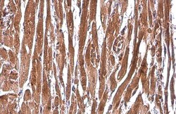
- Immunohistochemistry (Paraffin) analysis of ANP was performed in paraffin-embedded rat heart tissue using ANP Polyclonal Antibody (Product # PA5-29559) at a dilution of 1:500. Antigen Retrieval: Citrate buffer, pH 6.0, 15 min.
- Submitted by
- Invitrogen Antibodies (provider)
- Main image
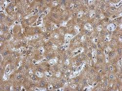
- Experimental details
- Immunohistochemical analysis of paraffin-embedded human hepatoma, using ANP (Product # PA5-29559) antibody at 1:500 dilution. Antigen Retrieval: EDTA based buffer, pH 8.0, 15 min.
Supportive validation
- Submitted by
- Invitrogen Antibodies (provider)
- Main image

- Experimental details
- Figure 1 Immunolocalization of ANP, NPR-A, and NPR-C in SGNs within the SG of P14 rats. Merge and single-channel images of cochlear sections triple labeled with antibodies against neural marker TUJ1 (green), ANP/NPR-A/NPR-C (red), and DAPI (blue). (a) ANP was predominantly immunoreactive in the perikarya of SGNs. NPR-A (b) and NPR-C (c) were predominantly immunoreactive in the plasma membrane and cytoplasm of SGNs and appeared more pronounced in the cellular membrane. Scale bars = 50 mu m.
- Submitted by
- Invitrogen Antibodies (provider)
- Main image
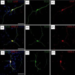
- Experimental details
- Figure 2 Immunolocalization of ANP, NPR-A, and NPR-C in dissociated SGNs from P3 rats. Merge and single-channel images of SG cells triple labeled with antibodies against TUJ1 (green), ANP/NPR-A/NPR-C (red), and DAPI (blue). The immunoreactivity of ANP (a), NPR-A (b), or NPR-C (c) was colocalized with beta -III tubulin-positive SGNs, respectively, distributed in the neuronal soma and neurites. Scale bars = 50 mu m.
- Submitted by
- Invitrogen Antibodies (provider)
- Main image
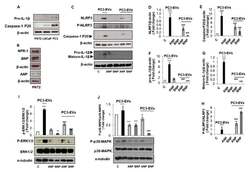
- Experimental details
- Figure 4 ANP and BNP counteract PC3-derived EVs-induced inflammasome activation in PNT2 cells. Non-cancerous PNT2 cells were grown for 24 h. Cell lysates were immunoblotted for pro- IL-1beta and mature caspase-1-p20 ( A ); NPR-1, ANP, and BNP ( B ). PNT2 cells were pre-incubated with ANP or BNP (1 uM) for 10 min and then treated with PC3-derived EVs (PC3-EVs) (100 ug/mL) for a 24 h. Cell lysate were immunoblotted for NLRP3 ( C , D ), phospho-NLRP3 (Ser295) ( C , H ), caspase-1 ( C , E ), IL-1beta ( C , F , G ), phospho-ERK 1/2 or ERK 1/2 ( I ) and phospho-p38-MAPK or p38-MAPK ( J ). beta-actin or alpha-tubulin were used as a loading control. Representative Western blots images are shown. Histograms represent densitometric quantification. All histograms indicate the mean +- SD of at least n = 3 independent experiments, each one tested in triplicate. ** p < 0.01, *** p < 0.001 vs. untreated PC3 cells. # p < 0.05, ### p < 0.001 vs. PC3-EVs treated PC3 cells.
- Submitted by
- Invitrogen Antibodies (provider)
- Main image

- Experimental details
- FIGURE 2 Immunolocalization of ANP, NPR-A and NPR-C in SGNs within cochlear SG from rats at P7. (A-C) Gross view of cryosectioned cochlear tissues triple-labeled with antibodies against neural marker beta-III Tubulin (TUJ1, green), ANP/NPR-A/NPR-C (red) and Hoechst (blue). (A 1 ,B 1 ,C 1 ) High-magnification views of the boxed region of (A-C) . (A 2 ,B 2 ,C 2 ) Higher-magnification images of the boxed region of (A 1 ,B 1 ,C 1 ) reveal that ANP/NPR-A/NPR-C is colocalized with TUJ1-positive SGNs, respectively. (A 2' ,B 2' ,C 2' ) Immunoreactivities of ANP, NPR-A, and NPR-C are present in the SGNs. Scale bars = 100 mum.
- Submitted by
- Invitrogen Antibodies (provider)
- Main image

- Experimental details
- FIGURE 2 Administration of recombinant vaspin protein attenuates cardiac hypertrophy and fibrosis in A-ZIP/F-1 lipoatrophic mice. Sixteen-week-old A-ZIP/F-1 mice were treated with recombinant vaspin protein (100 mug/mouse) by an osmatic pump for 8 weeks. (A) The ratio of heart weight/tibia length. (B,C) Representative images of whole hearts, and cardiac slides were stained with hematoxylin and eosin solution (B) , and quantitative analysis of average cardiomyocyte size (C) . Scale bar = 20 mum. (D,E) Western blot analysis of cardiac atrial natriuretic peptide (ANP) and B-type natriuretic peptide (BNP) (D) , and quantitative analysis of relative density of ANP/tubulin and BNP/tubulin (E) . (F,G) . Cardiac slides were stained with Sirius red solution (F) and quantitative analysis of relative density of collagen (G) . Scale bar = 40 mum. (H,I) Western blot analysis of cardiac TGF-beta1 and SMAD3 (H) and quantitative analysis of relative density of TGF-beta1/tubulin and SMAD3/tubulin (I) . Results are shown as mean +- SEM, and n = 8 mice/group. * p < 0.05, ** p < 0.01, *** p < 0.001.
- Submitted by
- Invitrogen Antibodies (provider)
- Main image

- Experimental details
- The protein level of RIP3 is elevated in hypertrophic hearts. (a) Representative Western blots and quantitation of RIP3 expression in heart tissue from patients with dilated cardiomyopathy (DCM) and normal controls. (b) (Up) Western blots of RIP3, ANP, beta -MHC, and GAPDH and (down) relative quantitation of RIP3 expression in heart tissue from rats after sham or AB operation. (c) (Up) Western blots of RIP3, ANP, beta -MHC, and GAPDH and (down) relative quantitation of RIP3 in NRCMs stimulated with Ang-II (1 mu M) or PBS. (d) (up) Western blots of RIP3, ANP, beta -MHC, and GAPDH and (down) relative quantitation of RIP3 in NRCMs stimulated with PE (50 mu M) or PBS. kDa: kilo-Dalton; * P < 0.05, ** P < 0.01, and *** P < 0.001.
 Explore
 Validate
 Learn
Learn Western blot