Explore
 Validate
 Learn
Learn Western blot
Antibody data
- Antibody Data
- Antigen structure
- References [1]
- Comments [0]
- Validations
- Western blot [1]
- Immunocytochemistry [1]
- Immunohistochemistry [5]
Submit
Validation data
Reference
Comment
Report error
- Product number
- HPA004872 - Provider product page

- Provider
- Atlas Antibodies
- Proper citation
- Atlas Antibodies Cat#HPA004872, RRID:AB_1079053
- Product name
- Anti-HGS
- Antibody type
- Polyclonal
- Reactivity
- Human
- Host
- Rabbit
- Conjugate
- Unconjugated
- Antigen sequence
LAEDIDPELARYLNRNYWEKKQEEARKSPTPSAPV
PLTEPAAQPGEGHAAPTNVVENPLPETDSQPIPPS
GGPFSEPQFHNGESEESHEQFLKALQNAVTTFVNR
MKSNHMRGRSITNDS- Isotype
- IgG
- Vial size
- 100 µl
- Storage
- Store at +4°C for short term storage. Long time storage is recommended at -20°C.
Submitted references T Cell Receptor (TCR)-induced Tyrosine Phosphorylation Dynamics Identifies THEMIS as a New TCR Signalosome Component
Brockmeyer C, Paster W, Pepper D, Tan C, Trudgian D, McGowan S, Fu G, Gascoigne N, Acuto O, Salek M
Journal of Biological Chemistry 2011 February;286(9):7535-7547
Journal of Biological Chemistry 2011 February;286(9):7535-7547
No comments: Submit comment
Supportive validation
- Submitted by
- Atlas Antibodies (provider)
- Enhanced method
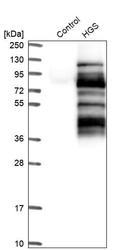
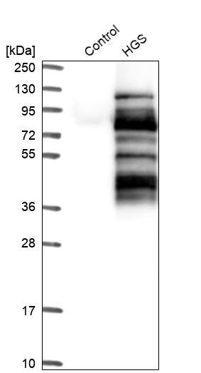
- Recombinant expression validation
- Main image
- Experimental details
- Western blot analysis in control (vector only transfected HEK293T lysate) and HGS over-expression lysate (Co-expressed with a C-terminal myc-DDK tag (~3.1 kDa) in mammalian HEK293T cells, LY401488).
Supportive validation
- Submitted by
- Atlas Antibodies (provider)
- Main image

- Experimental details
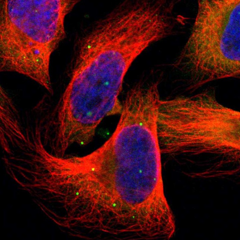
- Immunofluorescent staining of human cell line U-2 OS shows localization to cytosol & vesicles.
- Sample type
- HUMAN
Supportive validation
- Submitted by
- Atlas Antibodies (provider)
- Main image
- Experimental details
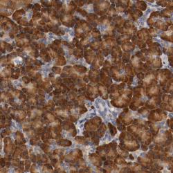
- Immunohistochemical staining of human pancreas shows strong cytoplasmic positivity in exocrine glandular cells.

- Submitted by
- Atlas Antibodies (provider)
- Main image
- Experimental details
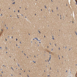
- Immunohistochemical staining of human cerebral cortex shows moderate positivity in neuropil.
- Sample type
- HUMAN

- Submitted by
- Atlas Antibodies (provider)
- Main image
- Experimental details
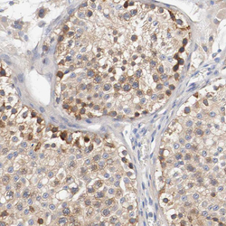
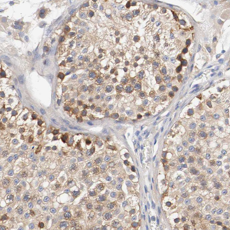
- Immunohistochemical staining of human testis shows moderate cytoplasmic positivity in cells in seminiferous ducts.
- Sample type
- HUMAN
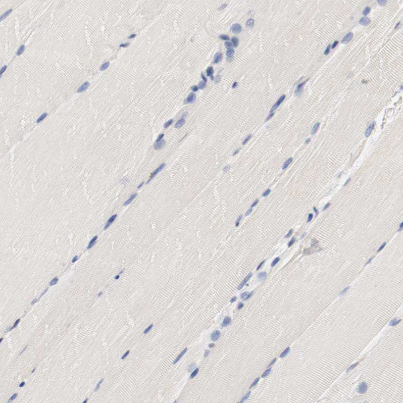
- Submitted by
- Atlas Antibodies (provider)
- Main image
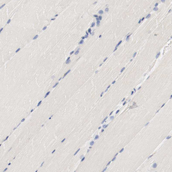
- Experimental details
- Immunohistochemical staining of human skeletal muscle shows no positivity in myocytes as expected.
- Sample type
- HUMAN
- Submitted by
- Atlas Antibodies (provider)
- Main image

- Experimental details
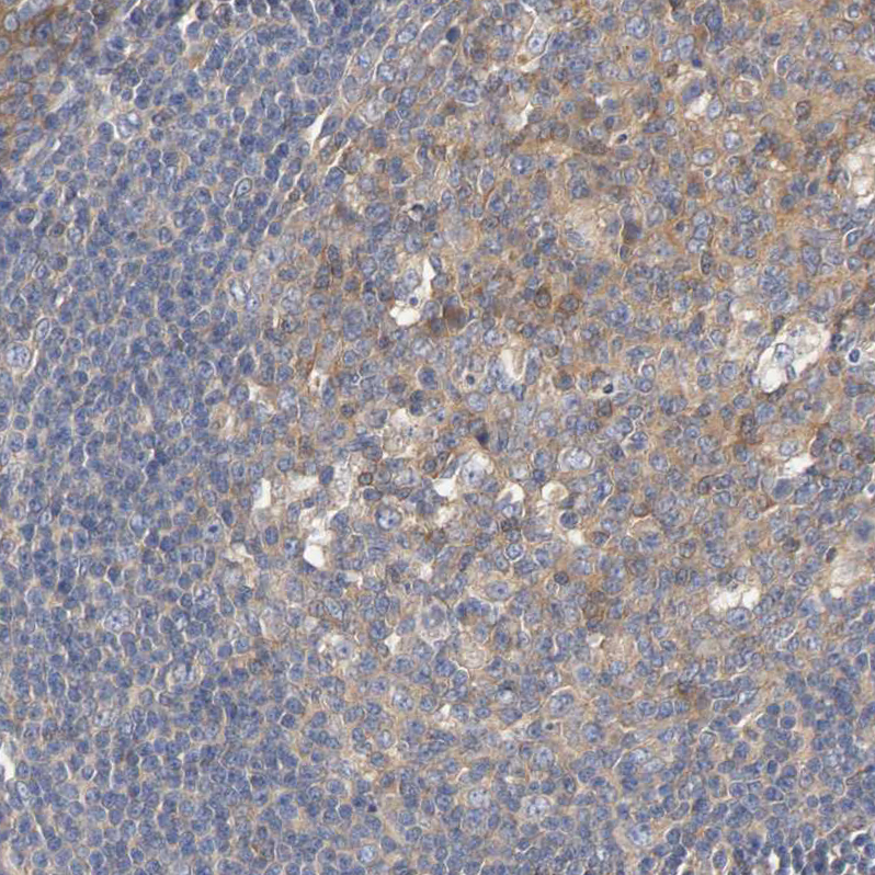
- Immunohistochemical staining of human tonsil shows moderate cytoplasmic positivity in germinal center cells.
- Sample type
- HUMAN