Explore
 Validate
 Learn
Learn Western blot
Antibody data
- Antibody Data
- Antigen structure
- References [1]
- Comments [0]
- Validations
- Western blot [1]
- Immunocytochemistry [2]
- Immunohistochemistry [1]
Submit
Validation data
Reference
Comment
Report error
- Product number
- MA1-036X - Provider product page

- Provider
- Invitrogen Antibodies
- Product name
- SLC2A5 Monoclonal Antibody (14C8)
- Antibody type
- Monoclonal
- Antigen
- Other
- Description
- This antibody requires overnight incubation in a Western blot for optimal performance. Do not use this antibody in a Fast Western blotting procedure.
- Reactivity
- Human, Mouse
- Host
- Mouse
- Isotype
- IgG
- Antibody clone number
- 14C8
- Vial size
- 20 µL
- Concentration
- 1 mg/mL
- Storage
- -20°C
Submitted references Analysis of glucose metabolism by (18)F-FDG-PET imaging and glucose transporter expression in a mouse model of intracerebral hemorrhage.
Han X, Ren H, Nandi A, Fan X, Koehler RC
Scientific reports 2021 May 25;11(1):10885
Scientific reports 2021 May 25;11(1):10885
No comments: Submit comment
Supportive validation
- Submitted by
- Invitrogen Antibodies (provider)
- Main image
- Experimental details
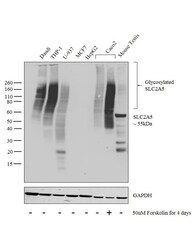
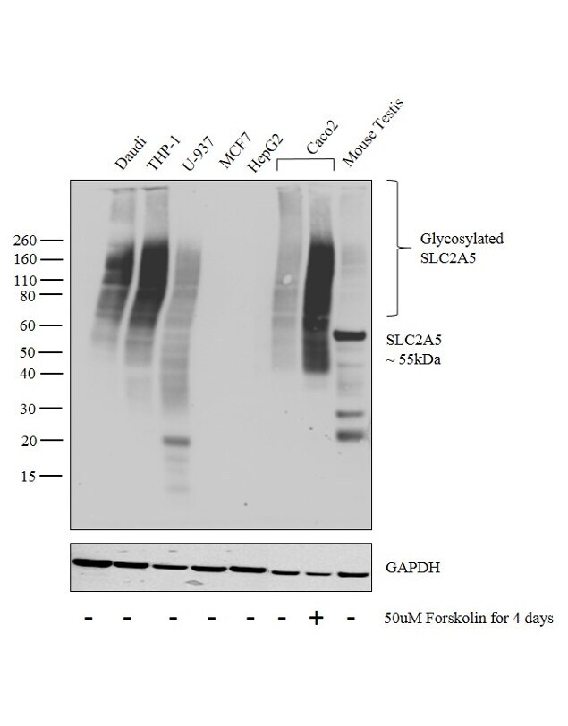
- Western blot analysis was performed on membrane enriched extracts (30 µg lysate) of Daudi (Lane 1), THP-1 (Lane 2), U-937 (Lane 3), MCF7 (Lane 4), HepG2 (Lane 5), Caco2 (Lane 6), Caco2 treated with Forskolin (50uM for 4 days) (Lane 7) and tissue extracts of Mouse Testis (Lane 8). The blot was probed with Anti-SLC2A5 Monoclonal Antibody (Product # MA1-036, 1:1000 dilution) and detected by chemiluminescence using Goat anti-Mouse IgG (H+L) Superclonal™ Secondary Antibody, HRP conjugate (Product # A28177, 0.25 µg/ml, 1:4000 dilution). High molecular weight corresponding to the glycosylated forms of SLC2A5 were observed in all the cell lines tested except MCF7 and HepG2. An 5kDa band corresponding to SLC2A5 was observed in Mouse Testis and the protein was also observed to be induced upon treatment of Caco2 cells with Forskolin.
Supportive validation
- Submitted by
- Invitrogen Antibodies (provider)
- Main image
- Experimental details
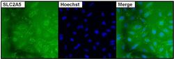
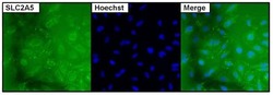
- Immunofluorescent analysis of SLC2A5 using anti-SLC2A5 monoclonal antibody (Product # MA1-036) (shown in green) in HeLa cells. Formalin fixed cells were permeabilized with 0.1% Triton X-100 in TBS for 10 minutes at room temperature. Cells were then blocked with 1% Blocker BSA (Product # 37525) for 15 minutes at room temperature. Cells were probed with a mouse monoclonal antibody recognizing SLC2A5 (Product # MA1-036), at a dilution of 1:100 for at least 1 hour at room temperature. Cells were then washed with PBS and incubated with DyLight 488 goat-anti-mouse secondary antibody (Product # 35503) at a dilution of 1:400 for 30 minutes at room temperature. Nuclei (blue) were stained with Hoechst 33342 dye (Product # 62249). Images were taken on a Thermo Scientific ArrayScan at 20X magnification.

- Submitted by
- Invitrogen Antibodies (provider)
- Main image
- Experimental details
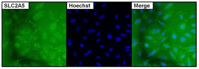
- Immunofluorescent analysis of SLC2A5 using anti-SLC2A5 monoclonal antibody (Product # MA1-036) (shown in green) in HeLa cells. Formalin fixed cells were permeabilized with 0.1% Triton X-100 in TBS for 10 minutes at room temperature. Cells were then blocked with 1% Blocker BSA (Product # 37525) for 15 minutes at room temperature. Cells were probed with a mouse monoclonal antibody recognizing SLC2A5 (Product # MA1-036), at a dilution of 1:100 for at least 1 hour at room temperature. Cells were then washed with PBS and incubated with DyLight 488 goat-anti-mouse secondary antibody (Product # 35503) at a dilution of 1:400 for 30 minutes at room temperature. Nuclei (blue) were stained with Hoechst 33342 dye (Product # 62249). Images were taken on a Thermo Scientific ArrayScan at 20X magnification.
Supportive validation
- Submitted by
- Invitrogen Antibodies (provider)
- Main image
- Experimental details
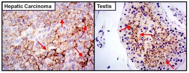
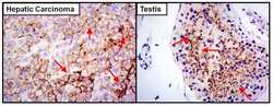
- Immunohistochemistry was performed on cancer biopsies of deparaffinized human testis and hepatic carcinoma tissues. To expose target proteins heat induced antigen retrieval was performed using 10mM sodium citrate (pH6.0) buffer for 20 minutes at 95°C. Following antigen retrieval tissues were blocked in 10% normal goat serum (Product # 31873) for 20 minutes at room temperature. Tissues were then probed at a dilution of 1:800 with a mouse monoclonal antibody recognizing SLC2A5 (Product # MA1-036) overnight at 4°C in a humidified chamber. Detection was performed using a goat anti-mouse HRP secondary antibody followed by colorimetric detection using DAB substrate. Tissues were counterstained with hematoxylin and prepped for mounting. Images were taken at 400X magnification. Results demonstrate membrane/cytoplasmic localization of SLC2A5.