 Explore
 Validate
 Learn
Learn Western blot
 ELISA
ELISAAntibody data
- Antibody Data
- Antigen structure
- References [1]
- Comments [0]
- Validations
- Western blot [2]
- Immunocytochemistry [1]
- Immunohistochemistry [2]
- Other assay [1]
Submit
Validation data
Reference
Comment
Report error
- Product number
- PA5-98154 - Provider product page

- Provider
- Invitrogen Antibodies
- Product name
- NKCC1 Polyclonal Antibody
- Antibody type
- Polyclonal
- Antigen
- Recombinant full-length protein
- Reactivity
- Human
- Host
- Rabbit
- Isotype
- IgG
- Vial size
- 100 µg
- Concentration
- 1 mg/mL
- Storage
- -20°C or -80°C if preferred
Submitted references Lack of NHE6 and Inhibition of NKCC1 Associated With Increased Permeability in Blood Labyrinth Barrier-Derived Endothelial Cell Layer.
Sekulic-Jablanovic M, Paproth J, Sgambato C, Albano G, Fuster DG, Bodmer D, Petkovic V
Frontiers in cellular neuroscience 2022;16:862119
Frontiers in cellular neuroscience 2022;16:862119
No comments: Submit comment
Supportive validation
- Submitted by
- Invitrogen Antibodies (provider)
- Main image

- Experimental details
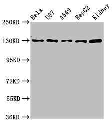
- Western Blot analysis of NKCC1 using a NKCC1 Polyclonal antibody (Product # PA5-98154) at a concentration of 6.8 µg/mL. Positive WB detected in: Hela whole cell lysate, U87 whole cell lysate, A549 whole cell lysate, HepG2 whole cell lysate, Mouse kidney tissue. A secondary Goat polyclonal antibody to rabbit IgG was applied at a 1:50,000 dilution. Observed band size: 132 kDa.
- Submitted by
- Invitrogen Antibodies (provider)
- Main image

- Experimental details
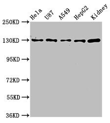
- Western Blot analysis of NKCC1 using a NKCC1 Polyclonal antibody (Product # PA5-98154) at a concentration of 6.8 µg/mL. Positive WB detected in: Hela whole cell lysate, U87 whole cell lysate, A549 whole cell lysate, HepG2 whole cell lysate, Mouse kidney tissue. A secondary Goat polyclonal antibody to rabbit IgG was applied at a 1:50,000 dilution. Observed band size: 132 kDa.
Supportive validation
- Submitted by
- Invitrogen Antibodies (provider)
- Main image
- Experimental details
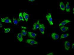
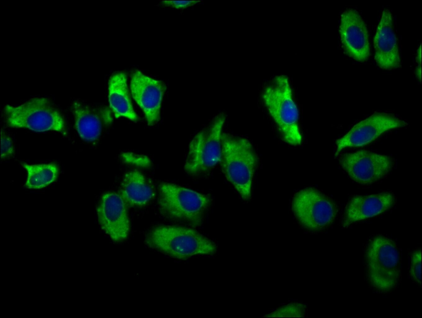
- Immunofluorescent analysis of NKCC1 in A549 cells using a NKCC1 polyclonal antibody (Product # PA5-98154) at a dilution of 1:100. The cells were fixed in 4% formaldehyde, permeabilized using 0.2% Triton X-100 and blocked in 10% normal Goat Serum. The cells were then incubated with the antibody overnight at 4°C. The secondary antibody was Alexa Fluor 488-congugated Goat Anti-Rabbit IgG(H+L). Cells were counter-stained with DAPI.
Supportive validation
- Submitted by
- Invitrogen Antibodies (provider)
- Main image
- Experimental details
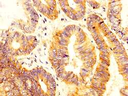
- Immunohistochemical analysis of NKCC1 in paraffin embedded human colon cancer using a NKCC1 polyclonal antibody (Product # PA5-98154) at a dilution of 1:300. After dewaxing and hydration, antigen retrieval was mediated by high pressure in a citrate buffer (pH 6.0). Section was blocked with 10% normal goat serum 30min at RT. Then primary antibody (1% BSA) was incubated at 4°C overnight. The primary is detected by a biotinylated secondary antibody and visualized using an HRP conjugated SP system.

- Submitted by
- Invitrogen Antibodies (provider)
- Main image

- Experimental details
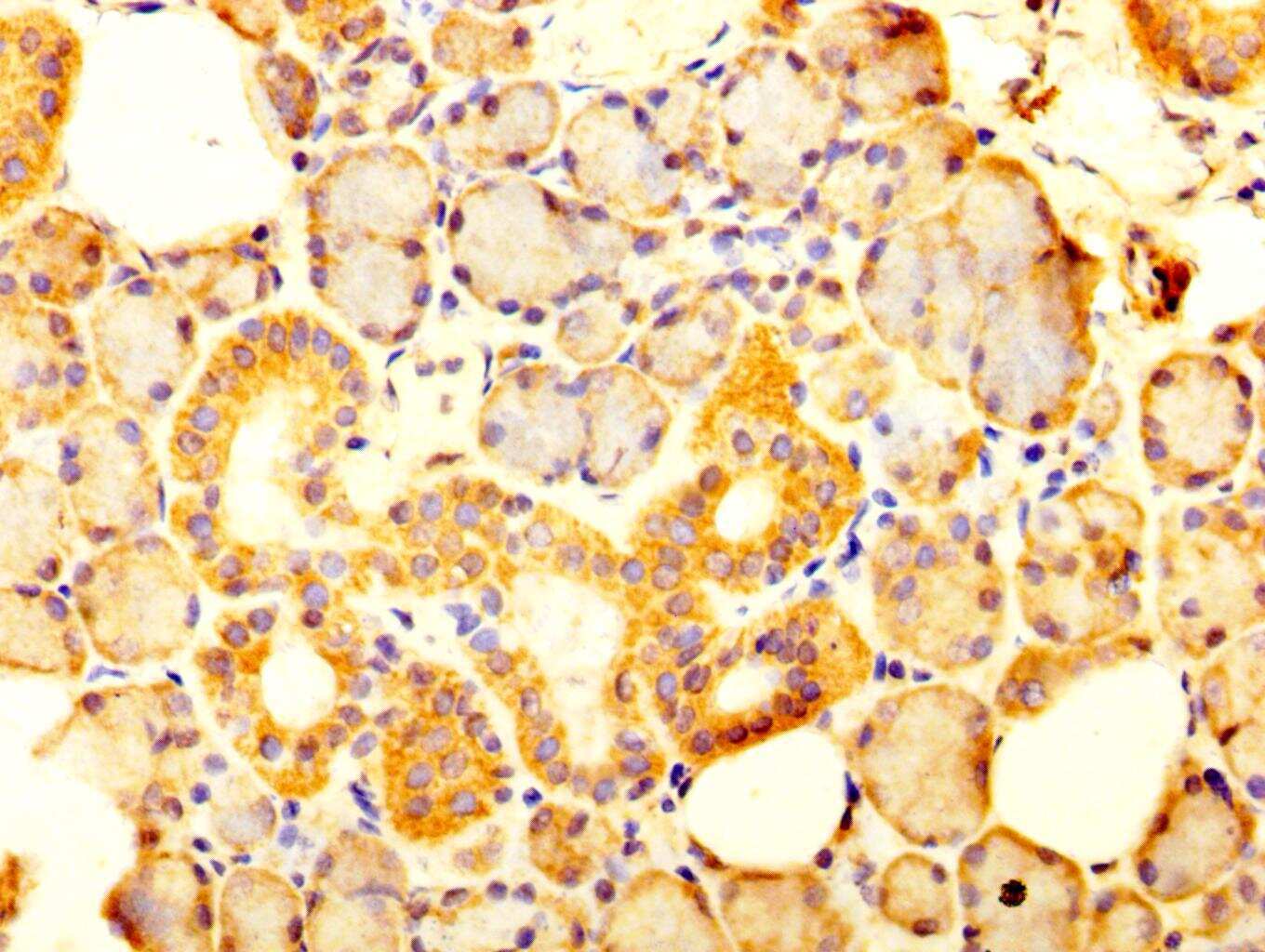
- Immunohistochemical analysis of NKCC1 in paraffin embedded human salivary gland tissue using a NKCC1 polyclonal antibody (Product # PA5-98154) at a dilution of 1:300. After dewaxing and hydration, antigen retrieval was mediated by high pressure in a citrate buffer (pH 6.0). Section was blocked with 10% normal goat serum 30min at RT. Then primary antibody (1% BSA) was incubated at 4°C overnight. The primary is detected by a biotinylated secondary antibody and visualized using an HRP conjugated SP system.
Supportive validation
- Submitted by
- Invitrogen Antibodies (provider)
- Main image
- Experimental details
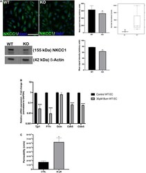
- FIGURE 4 NKCC1 inhibition influences endothelial permeability. (A, top left panel) endothelial cells immunostained with anti-NKCC1 antibodies show homogenous protein expression in a primary culture of wildtype (WT) and Nhe6 -knockout endothelial cells (KO). (A, middle panel) NKCC1 immunofluorescence signal quantification. Samples from five mice were pooled per phenotype and experiment; ( n = 3); Scale bar, 50 mum. Data represent the mean (SD); ( n = 3); ** P < 0.01. (A, top right panel) In Nhe6 -knockout cells (KO), NKCC1 expression was downregulated, compared to WT cells. Data represent the mean +- SD; ( n = 3); * P < 0.05. (A, bottom left panel) Representative western blot image showing decreased NKCC1 protein presence in Nhe6 -knockout EC cell lysate. (A, bottom right panel) western bolt signal quantification. Each endothelial cell culture was obtained from three mice per phenotype and experiment, and individual experiments performed three times ( n = 3); Data represent the mean (SD); ** P < 0.01. (B) Relative mRNA expression of the junctional genes after inhibiting NKCC1 with 30 muM bumetanide (bum) in WT endothelial cells (EC). Error bars represent the (SD); ( n = 3); **** P < 0.0001. (C) After inhibiting NKCC1 in WT ECs with 30 muM bumetanide (30 muM), permeability increased compared to untreated control cells (CTRL). Data represent the mean (SD); ( n = 3). * P < 0.05.
