 Explore
 Validate
 Learn
Learn Western blot
Antibody data
- Antibody Data
- Antigen structure
- References [4]
- Comments [0]
- Validations
- Western blot [3]
- Immunocytochemistry [2]
- Immunohistochemistry [2]
- Flow cytometry [1]
- Other assay [1]
Submit
Validation data
Reference
Comment
Report error
- Product number
- 42-2800 - Provider product page

- Provider
- Invitrogen Antibodies
- Product name
- Pannexin 2 Polyclonal Antibody
- Antibody type
- Polyclonal
- Antigen
- Synthetic peptide
- Description
- Applications Reported: YB5.B8 has been reported for use in flow cytometric analysis, immunoprecipitation, and immunohistochemical staining of frozen tissue sections.
- Reactivity
- Human, Mouse, Rat
- Host
- Rabbit
- Isotype
- IgG
- Vial size
- 100 µg
- Concentration
- 0.25 mg/mL
- Storage
- -20°C
Submitted references Synaptic Scaffolds, Ion Channels and Polyamines in Mouse Photoreceptor Synapses: Anatomy of a Signaling Complex.
A Potential Compensatory Role of Panx3 in the VNO of a Panx1 Knock Out Mouse Model.
Membrane-associated guanylate kinase scaffolds organize a horizontal cell synaptic complex restricted to invaginating contacts with photoreceptors.
Pannexins are new molecular candidates for assembling gap junctions in the cochlea.
Vila A, Shihabeddin E, Zhang Z, Santhanam A, Ribelayga CP, O'Brien J
Frontiers in cellular neuroscience 2021;15:667046
Frontiers in cellular neuroscience 2021;15:667046
A Potential Compensatory Role of Panx3 in the VNO of a Panx1 Knock Out Mouse Model.
Whyte-Fagundes P, Kurtenbach S, Zoidl C, Shestopalov VI, Carlen PL, Zoidl G
Frontiers in molecular neuroscience 2018;11:135
Frontiers in molecular neuroscience 2018;11:135
Membrane-associated guanylate kinase scaffolds organize a horizontal cell synaptic complex restricted to invaginating contacts with photoreceptors.
Vila A, Whitaker CM, O'Brien J
The Journal of comparative neurology 2017 Mar 1;525(4):850-867
The Journal of comparative neurology 2017 Mar 1;525(4):850-867
Pannexins are new molecular candidates for assembling gap junctions in the cochlea.
Tang W, Ahmad S, Shestopalov VI, Lin X
Neuroreport 2008 Aug 27;19(13):1253-7
Neuroreport 2008 Aug 27;19(13):1253-7
No comments: Submit comment
Supportive validation
- Submitted by
- Invitrogen Antibodies (provider)
- Main image

- Experimental details
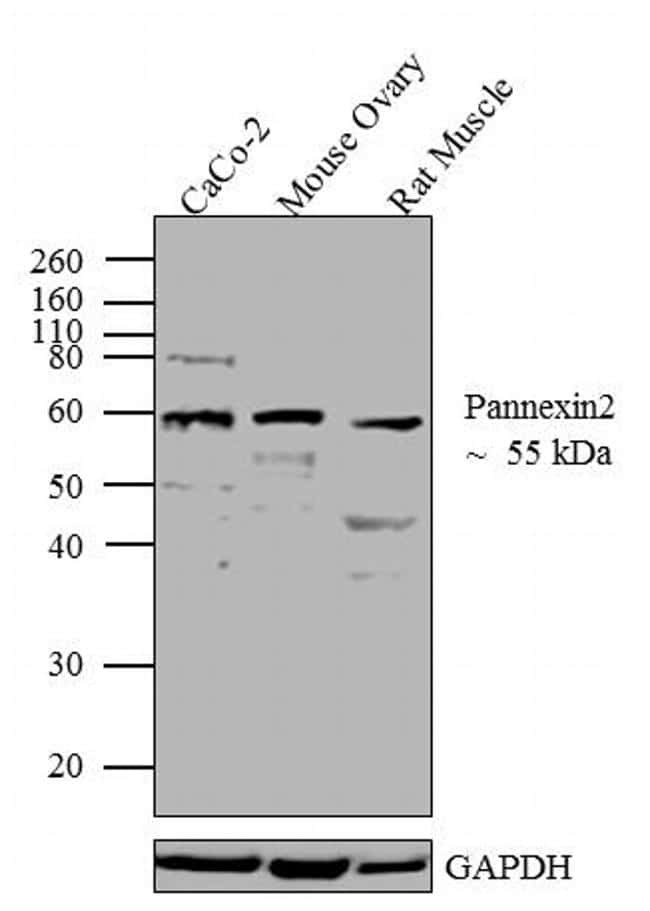
- Western blot analysis was performed on whole cell extracts (20 µg lysate) of CaCo-2 (Lane 1), Mouse Ovary (Lane 2) and Rat Muscle (lane 3). The blots were probed with Anti-Pannexin 2 Rabbit Polyclonal Antibody (Product # 42-2800, 0.5-2 µg/mL) and detected by chemiluminescence using Goat anti-Rabbit IgG (H+L) Superclonal™ Secondary Antibody, HRP conjugate (Product # A27036, 0.4 µg/mL, 1:2500 dilution). A 55 kDa band corresponding to Pannexin 2 was observed across cell lines and tissues tested. Known quantity of protein samples were electrophoresed using Novex® NuPAGE® 12 % Bis-Tris gel (Product # NP0342BOX), XCell SureLock™ Electrophoresis System (Product # EI0002) and Novex® Sharp Pre-Stained Protein Standard (Product # LC5800). Resolved proteins were then transferred onto a nitrocellulose membrane with iBlot® 2 Dry Blotting System (Product # IB21001). The membrane was probed with the relevant primary and secondary Antibody using iBind™ Flex Western Starter Kit (Product # SLF2000S). Chemiluminescent detection was performed using Pierce™ ECL Western Blotting Substrate (Product # 32106).

- Submitted by
- Invitrogen Antibodies (provider)
- Main image

- Experimental details
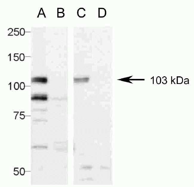
- Western blot analysis of (A & C) pannexin 2-GFP- transfected C6 cells and (B & D) untransfected C6 cells using (A & B) Zymed Rb anti-Pannexin 2 (C-term) (Product # 42-2800) and anti-GFP (C & D). Image courtesy of Dr. James Nagy, University of Manitoba, Winnipeg, Canada.
- Submitted by
- Invitrogen Antibodies (provider)
- Main image

- Experimental details
- Western blot analysis was performed on whole cell extracts (20 µg lysate) of CaCo-2 (Lane 1), Mouse Ovary (Lane 2) and Rat Muscle (lane 3). The blots were probed with Anti-Pannexin 2 Rabbit Polyclonal Antibody (Product # 42-2800, 0.5-2 µg/mL) and detected by chemiluminescence using Goat anti-Rabbit IgG (H+L) Superclonal™ Secondary Antibody, HRP conjugate (Product # A27036, 0.4 µg/mL, 1:2500 dilution). A 55 kDa band corresponding to Pannexin 2 was observed across cell lines and tissues tested. Known quantity of protein samples were electrophoresed using Novex® NuPAGE® 12 % Bis-Tris gel (Product # NP0342BOX), XCell SureLock™ Electrophoresis System (Product # EI0002) and Novex® Sharp Pre-Stained Protein Standard (Product # LC5800). Resolved proteins were then transferred onto a nitrocellulose membrane with iBlot® 2 Dry Blotting System (Product # IB21001). The membrane was probed with the relevant primary and secondary Antibody using iBind™ Flex Western Starter Kit (Product # SLF2000S). Chemiluminescent detection was performed using Pierce™ ECL Western Blotting Substrate (Product # 32106).
Supportive validation
- Submitted by
- Invitrogen Antibodies (provider)
- Main image

- Experimental details
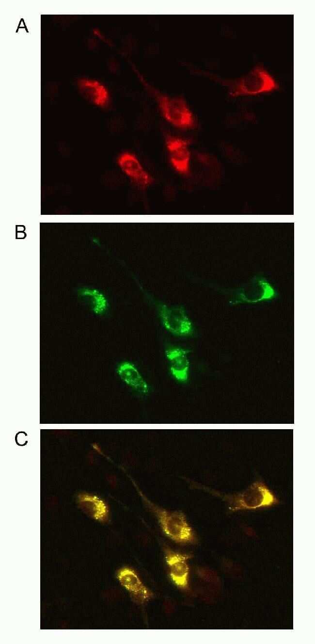
- Indirect immunofluorescence staining of GFP-pannexin-2-transfected C6 cells using (A) Zymed (R) Rb anti-Pannexin 2 (C-term) (Product # 42-2800). GFP fluorescence is shown in (B), and the images have been merged in (C). Image courtesy of James Nagy, Ph.D., University of Manitoba, Canada.
- Submitted by
- Invitrogen Antibodies (provider)
- Main image

- Experimental details
- Immunofluorescence analysis of Pannexin 2 was done on 90% confluent log phase Caco2 cells. The cells were fixed with 4% paraformaldehyde for 10 minutes, permeabilized with 0.1% Triton™ X-100 for 10 minutes, and blocked with 1% BSA for 1 hour at room temperature. The cells were labeled with Pannexin 2 Rabbit Polyclonal Antibody (Product # 42-2800) at 2 µg/mL in 0.1% BSA and incubated for 3 hours at room temperature and then labeled with Goat anti-Rabbit IgG (H+L) Superclonal™ Secondary Antibody, Alexa Fluor® 488 conjugate (Product # A27034) at a dilution of 1:2000 for 45 minutes at room temperature (Panel a: green). Nuclei (Panel b: blue) were stained with SlowFade® Gold Antifade Mountant with DAPI (Product # S36938). F-actin (Panel c: red) was stained with Alexa Fluor® 555 Rhodamine Phalloidin (Product # R415, 1:300). Panel d is a merged image showing punctuated membranous localization. Panel e is a no primary antibody control. The images were captured at 60X magnification.

Supportive validation
- Submitted by
- Invitrogen Antibodies (provider)
- Main image
- Experimental details
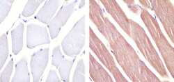
- Immunohistochemistry analysis of Pannexin 2 showing staining in the cytoplasm of paraffin-embedded mouse skeletal muscle tissue (right) compared to a negative control without primary antibody (left). To expose target proteins, antigen retrieval was performed using 10mM sodium citrate (pH 6.0), microwaved for 8-15 min. Following antigen retrieval, tissues were blocked in 3% H2O2-methanol for 15 min at room temperature, washed with ddH2O and PBS, and then probed with a Anti- Pannexin 2 Polyclonal Antibody (Product # 42-2800) diluted in 3% BSA-PBS at a dilution of 1:20 overnight at 4ºC in a humidified chamber. Tissues were washed extensively in PBST and detection was performed using an HRP-conjugated secondary antibody followed by colorimetric detection using a DAB kit. Tissues were counterstained with hematoxylin and dehydrated with ethanol and xylene to prep for mounting.

- Submitted by
- Invitrogen Antibodies (provider)
- Main image

- Experimental details
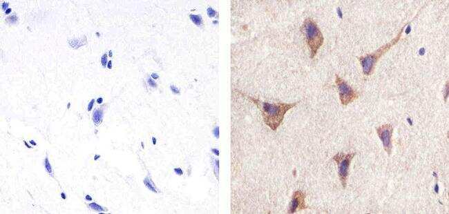
- Immunohistochemistry analysis of Pannexin 2 showing staining in the cytoplasm of paraffin-embedded human brain tissue (right) compared to a negative control without primary antibody (left). To expose target proteins, antigen retrieval was performed using 10mM sodium citrate (pH 6.0), microwaved for 8-15 min. Following antigen retrieval, tissues were blocked in 3% H2O2-methanol for 15 min at room temperature, washed with ddH2O and PBS, and then probed with a Anti-Pannexin 2 Polyclonal Antibody (Product # 42-2800) diluted in 3% BSA-PBS at a dilution of 1:100 overnight at 4ºC in a humidified chamber. Tissues were washed extensively in PBST and detection was performed using an HRP-conjugated secondary antibody followed by colorimetric detection using a DAB kit. Tissues were counterstained with hematoxylin and dehydrated with ethanol and xylene to prep for mounting.
Supportive validation
- Submitted by
- Invitrogen Antibodies (provider)
- Main image

- Experimental details
- Flow cytometry analysis of Pannexin 2 was done on SH-SY5Y cells. Cells were fixed with 70% ethanol for 10 minutes, permeabilized with 0.25% Triton™ X-100 for 20 minutes, and blocked with 5% BSA for 30 minutes at room temperature. Cells were labeled with Pannexin 2 Rabbit Polyclonal Antibody (422800, red histogram) or with rabbit isotype control (yellow histogram) at 3-5 ug/million cells in 2.5% BSA. After incubation at room temperature for 2 hours, the cells were labeled with Alexa Fluor® 488 Goat Anti-Rabbit Secondary Antibody (A11008) at a dilution of 1:400 for 30 minutes at room temperature. The representative 10,000 cells were acquired and analyzed for each sample using an Attune® Acoustic Focusing Cytometer. The purple histogram represents unstained control cells and the green histogram represents no-primary-antibody control..

Supportive validation
- Submitted by
- Invitrogen Antibodies (provider)
- Main image

- Experimental details
- FIGURE 5 Pannexin labeling in mouse retina. (A) Immunostaining for Pannexin 1 in mouse retina revealed strong labeling in the outer plexiform layer and weaker labeling in the inner nuclear layer and ganglion cell layer. Three mum confocal stack. Labels for retinal nuclear layers are as in Figure 1 . (B) Immunostaining for Pannexin 2 displayed strong labeling in the OPL and weaker labeling in the INL and photoreceptor layer. Three mum confocal stack. (C) Average mRNA expression levels of Pannexin1 and Pannexin2 in mouse retina from single-cell transcriptome data. In the OPL, the most prominent expression of Pannexin1 is in Off cone bipolar cells, with very weak expression in horizontal cells. Pannexin2 expression is largely absent from horizontal cells, but weakly present in On and Off bipolar cells. Data adapted from .
