 Explore
 Validate
 Learn
LearnMAB3024
antibody from R&D Systems
Targeting: KEAP1
INrf2, KIAA0132, KLHL19, MGC10630, MGC1114, MGC20887, MGC4407, MGC9454
 Western blot
Antibody data
- Antibody Data
- Antigen structure
- References [2]
- Comments [0]
- Validations
- Western blot [2]
Submit
Validation data
Reference
Comment
Report error
- Product number
- MAB3024 - Provider product page

- Provider
- R&D Systems
- Product name
- Human/Mouse/Rat Keap1 Antibody
- Antibody type
- Monoclonal
- Description
- Protein A or G purified from hybridoma culture supernatant. Detects human, mouse and rat Keap1 in Western blots.
- Reactivity
- Human, Mouse, Rat
- Host
- Mouse
- Conjugate
- Unconjugated
- Antigen sequence
Q14145- Isotype
- IgG
- Antibody clone number
- 333116
- Vial size
- 100 ug
- Concentration
- LYOPH
- Storage
- Use a manual defrost freezer and avoid repeated freeze-thaw cycles. 12 months from date of receipt, -20 to -70 °C as supplied. 1 month, 2 to 8 °C under sterile conditions after reconstitution. 6 months, -20 to -70 °C under sterile conditions after reconstitution.
Submitted references Nrf2-Keap1 pathway promotes cell proliferation and diminishes ferroptosis.
Deficiency in the nuclear factor E2-related factor-2 transcription factor results in impaired adipogenesis and protects against diet-induced obesity.
Fan Z, Wirth AK, Chen D, Wruck CJ, Rauh M, Buchfelder M, Savaskan N
Oncogenesis 2017 Aug 14;6(8):e371
Oncogenesis 2017 Aug 14;6(8):e371
Deficiency in the nuclear factor E2-related factor-2 transcription factor results in impaired adipogenesis and protects against diet-induced obesity.
Pi J, Leung L, Xue P, Wang W, Hou Y, Liu D, Yehuda-Shnaidman E, Lee C, Lau J, Kurtz TW, Chan JY
The Journal of biological chemistry 2010 Mar 19;285(12):9292-300
The Journal of biological chemistry 2010 Mar 19;285(12):9292-300
No comments: Submit comment
Supportive validation
- Submitted by
- R&D Systems (provider)
- Main image
- Experimental details
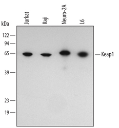
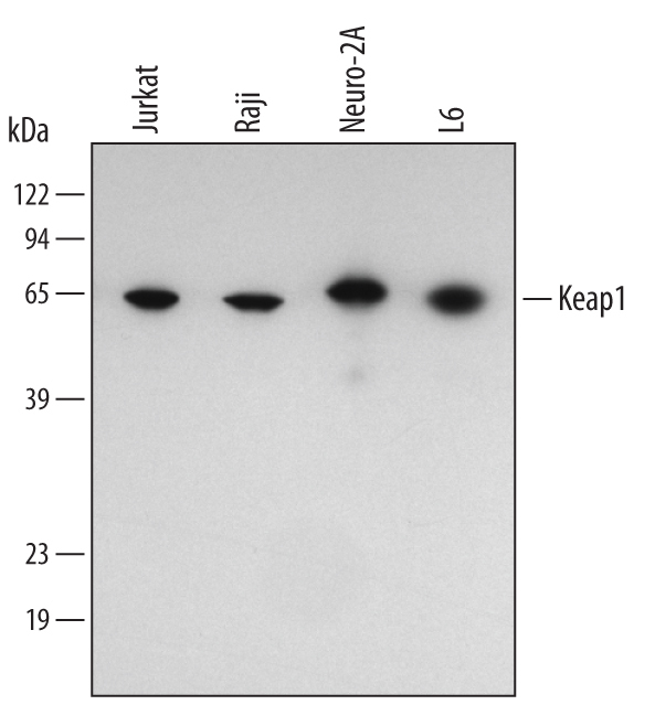
- Detection of Human/Mouse/Rat Keap1 by Western Blot. Western blot shows lysates of Jurkat human acute T cell leukemia cell line, Raji human Burkitt's lymphoma cell line, Neuro-2A mouse neuroblastoma cell line, and L6 rat myoblast cell line. PVDF membrane was probed with 1 µg/mL of Human/Mouse/Rat Keap1 Monoclonal Antibody (Catalog # MAB3024) followed by HRP-conjugated Anti-Mouse IgG Secondary Antibody (Catalog # HAF007). A specific band was detected for Keap1 at approximately 66 kDa (as indicated). This experiment was conducted under reducing conditions and using Immunoblot Buffer Group 4.
- Submitted by
- R&D Systems (provider)
- Main image
- Experimental details
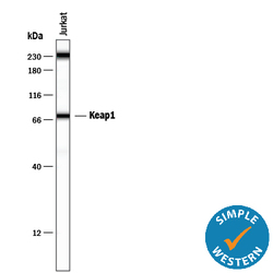
- Detection of Human Keap1 by Simple WesternTM. Simple Western lane view shows lysates of Jurkat human acute T cell leukemia cell line, loaded at 0.2 mg/mL. A specific band was detected for Keap1 at approximately 75 kDa (as indicated) using 20 µg/mL of Mouse Anti-Human/Mouse/Rat Keap1 Monoclonal Antibody (Catalog # MAB3024). This experiment was conducted under reducing conditions and using the 12-230 kDa separation system. Non-specific interaction with the 230 kDa Simple Western standard may be seen with this antibody.
