Explore
 Validate
 Learn
Learn Western blot
Antibody data
- Antibody Data
- Antigen structure
- References [3]
- Comments [0]
- Validations
- Western blot [3]
- Immunohistochemistry [1]
Submit
Validation data
Reference
Comment
Report error
- Product number
- GTX105316 - Provider product page

- Provider
- GeneTex
- Proper citation
- GeneTex Cat#GTX105316, RRID:AB_1951878
- Product name
- C1 inhibitor antibody [N1C3]
- Antibody type
- Polyclonal
- Reactivity
- Human, Bovine
- Host
- Rabbit
Submitted references Generation of α-1,3-galactosyltransferase knocked-out transgenic cloned pigs with knocked-in five human genes.
Aberrant glycosylation of plasma proteins in severe preeclampsia promotes monocyte adhesion.
Differential gene expression of serine protease inhibitors in bovine ovarian follicle: possible involvement in follicular growth and atresia.
Kwon DJ, Kim DH, Hwang IS, Kim DE, Kim HJ, Kim JS, Lee K, Im GS, Lee JW, Hwang S
Transgenic research 2017 Feb;26(1):153-163
Transgenic research 2017 Feb;26(1):153-163
Aberrant glycosylation of plasma proteins in severe preeclampsia promotes monocyte adhesion.
Flood-Nichols SK, Kazanjian AA, Tinnemore D, Gafken PR, Ogata Y, Napolitano PG, Stallings JD, Ippolito DL
Reproductive sciences (Thousand Oaks, Calif.) 2014 Feb;21(2):204-14
Reproductive sciences (Thousand Oaks, Calif.) 2014 Feb;21(2):204-14
Differential gene expression of serine protease inhibitors in bovine ovarian follicle: possible involvement in follicular growth and atresia.
Hayashi KG, Ushizawa K, Hosoe M, Takahashi T
Reproductive biology and endocrinology : RB&E 2011 May 27;9:72
Reproductive biology and endocrinology : RB&E 2011 May 27;9:72
No comments: Submit comment
Supportive validation
- Submitted by
- GeneTex (provider)
- Main image
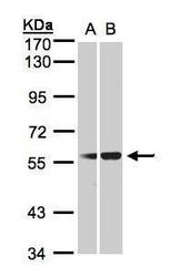
- Experimental details
- Sample(30 ug whole cell lysate)A:293TB:HeLa S3(GTX14654)7.5% SDS PAGEGTX105316 diluted at 1:1000
- Validation comment
- WB
- Submitted by
- GeneTex (provider)
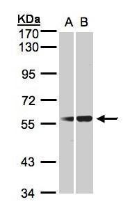
- Main image

- Experimental details
- Various whole cell extracts (30 ?g) were separated by 7.5% SDS-PAGE, and the membrane was blotted with C1 inhibitor antibody [N1C3] (GTX105316) diluted at 1:1000. The HRP-conjugated anti-rabbit IgG antibody (GTX213110-01) was used to detect the primary antibody.

- Submitted by
- GeneTex (provider)
- Main image
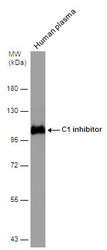
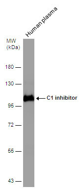
- Experimental details
- Human tissue extract (30 ?g) was separated by 7.5% SDS-PAGE, and the membrane was blotted with C1 inhibitor antibody [N1C3] (GTX105316) diluted at 1:5000. The HRP-conjugated anti-rabbit IgG antibody (GTX213110-01) was used to detect the primary antibody.
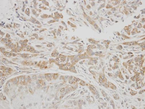
Supportive validation
- Submitted by
- GeneTex (provider)
- Main image

- Experimental details
- Immunohistochemical analysis of paraffin-embedded A549 xenograft, using C1 inhibitor(GTX105316) antibody at 1:300 dilution.