 Explore
 Validate
 Learn
LearnPA5-88997
antibody from Invitrogen Antibodies
Targeting: NLRC4
CARD12, CLAN, CLAN1, CLANA, CLANB, CLANC, CLAND, CLR2.1, ipaf
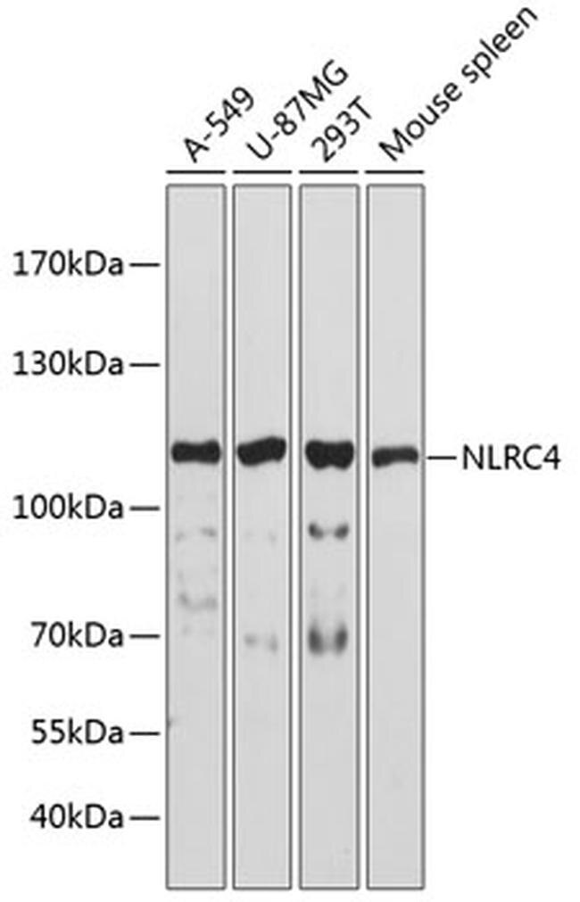
 Western blot
Antibody data
- Antibody Data
- Antigen structure
- References [2]
- Comments [0]
- Validations
- Western blot [1]
- Immunocytochemistry [3]
- Immunohistochemistry [4]
- Other assay [1]
Submit
Validation data
Reference
Comment
Report error
- Product number
- PA5-88997 - Provider product page

- Provider
- Invitrogen Antibodies
- Product name
- NLRC4 Polyclonal Antibody
- Antibody type
- Polyclonal
- Antigen
- Recombinant full-length protein
- Description
- For reconstitution, we recommend adding 100 µL distilled water to a final antibody concentration of about 1 mg/mL. To use this carrier-free antibody for conjugation experiments, we strongly recommend performing another round of desalting. (Zeba Spin Desalting Columns, 7KMWCO, 0.5 mL, Product # 89882)
- Reactivity
- Human, Mouse
- Host
- Rabbit
- Isotype
- IgG
- Vial size
- 100 µL
- Concentration
- 1.24 mg/mL
- Storage
- -20° C, Avoid Freeze/Thaw Cycles
Submitted references A role for the NLRC4 inflammasome in premature rupture of membrane.
oprC Impairs Host Defense by Increasing the Quorum-Sensing-Mediated Virulence of Pseudomonas aeruginosa.
Zhu J, Ma C, Zhu L, Li J, Peng F, Huang L, Luan X
PloS one 2020;15(8):e0237847
PloS one 2020;15(8):e0237847
oprC Impairs Host Defense by Increasing the Quorum-Sensing-Mediated Virulence of Pseudomonas aeruginosa.
Gao P, Guo K, Pu Q, Wang Z, Lin P, Qin S, Khan N, Hur J, Liang H, Wu M
Frontiers in immunology 2020;11:1696
Frontiers in immunology 2020;11:1696
No comments: Submit comment
Supportive validation
- Submitted by
- Invitrogen Antibodies (provider)
- Main image

- Experimental details
- Western blot analysis of extracts of various cell lines, using NLRC4 Polyclonal antibody (Product # PA5-88997) at 1:3000 dilution. Secondary antibody: HRP Goat Anti-Rabbit IgG (H+L) at 1:10000 dilution. Lysates/proteins: 25ug per lane. Blocking buffer: 3% nonfat dry milk in TBST. Exposure time: 60s.
Supportive validation
- Submitted by
- Invitrogen Antibodies (provider)
- Main image

- Experimental details
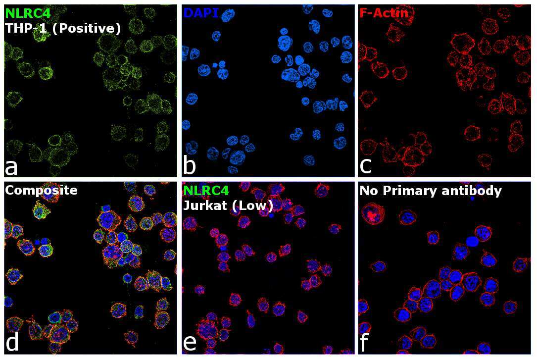
- Immunocytochemistry-Immunofluorescence analysis of NLRC4 was performed in THP-1 cells using NLRC4 Polyclonal Antibody (Product # PA5-88997).

- Submitted by
- Invitrogen Antibodies (provider)
- Main image

- Experimental details
- Immunofluorescence analysis of NLRC4 (NLR family CARD domain-containing protein 4) was performed using THP-1 and Jurkat cells. The cells were fixed with 4% paraformaldehyde for 10 minutes, permeabilized with 0.1% Triton™ X-100 for 10 minutes, and blocked with 2% BSA for 45 minutes at room temperature. The cells were labeled with NLRC4 Polyclonal Antibody (Product # PA5-88997) at 1:100 in 0.1% BSA, incubated at 4 degree celsius overnight and then labeled with Donkey anti-Rabbit IgG (H+L) Highly Cross-Adsorbed Secondary Antibody, Alexa Fluor Plus 488 (Product # A32790), (1:2500 dilution), for 45 minutes at room temperature (Panel a: Green). Nuclei (Panel b: Blue) were stained with ProLong™ Diamond Antifade Mountant with DAPI (Product # P36962). F-actin (Panel c: Red) was stained with Rhodamine Phalloidin (Product # R415, 1:300). Panel d represents the merged image showing cytoplasmic localization of NLRC4 in THP-1 and low levels of expression of the same in Jurkat (Panel e). Panel f represents control cells with no primary antibody to assess background. The images were captured at 60X magnification.
- Submitted by
- Invitrogen Antibodies (provider)
- Main image

- Experimental details
- Knockdown of NLRC4 was achieved by transfecting THP-1 cells with NLRC4 specific siRNA (Silencer® select Product # s33828, s33829). Immunofluorescence analysis was performed on untransfected THP-1 cells (panel a-d), transfected with non-specific scrambled siRNA (panels e-h) and transfected with NLRC4 specific siRNA (panel i-l). Cells were fixed, permeabilized, and labelled with NLRC4 Polyclonal Antibody (Product # PA5-88997, 1:100) followed by Donkey anti-Rabbit IgG (H+L) Highly Cross-Adsorbed Secondary Antibody, Alexa Fluor Plus 488 (Product # A32790), (1:2500 dilution). Nuclei (blue) were stained using ProLong™ Diamond Antifade Mountant with DAPI (Product # P36962), and Rhodamine Phalloidin (Product # R415, 1:300) was used for cytoskeletal F-actin (red) staining. Reduction in specific signal was observed upon siRNA mediated knockdown (panel i,l) (Green) confirming specificity of the antibody to NLRC4. The Images were captured at 60X magnification.

Supportive validation
- Submitted by
- Invitrogen Antibodies (provider)
- Main image

- Experimental details
- Immunohistochemistry analysis of NLRC4 in paraffin-embedded human lung using NLRC4 Polyclonal Antibody (Product # PA5-88997) at a dilution of 1:200.

- Submitted by
- Invitrogen Antibodies (provider)
- Main image

- Experimental details
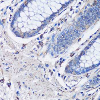
- Immunohistochemistry analysis of NLRC4 in paraffin-embedded human colon using NLRC4 Polyclonal Antibody (Product # PA5-88997) at a dilution of 1:100.
- Submitted by
- Invitrogen Antibodies (provider)
- Main image

- Experimental details
- Immunohistochemistry analysis of NLRC4 in paraffin-embedded human gastric cancer using NLRC4 Polyclonal Antibody (Product # PA5-88997) at a dilution of 1:100.

- Submitted by
- Invitrogen Antibodies (provider)
- Main image

- Experimental details
- Immunohistochemistry-Immunofluorescence analysis of NLRC4 was performed in mouse lung tissue using NLRC4 Polyclonal Antibody (Product # PA5-88997) at a dilution of 1:100. Blue: DAPI for nuclear staining.

Supportive validation
- Submitted by
- Invitrogen Antibodies (provider)
- Main image

- Experimental details
- Fig 2 Increased activation of NLRC4 inflammasomes in placenta and membranes of patients with TPROM. (A) RT-PCR analysis of NLRC4 at mRNA level in fetal membrane; (B) NLRC4 immunostaining in fetal membrane was shown, bar = 100 mum; (C) Relative mRNA level of NLRC4 in placenta; (D) Representative IHC staining images of placenta, bar = 100 mum; (E-H) mRNA abundance of inflammasome components and NOD2 in fetal membranes, *P
