Explore
 Validate
 Learn
Learn Western blot
 Immunocytochemistry
Antibody data
- Antibody Data
- Antigen structure
- References [5]
- Comments [0]
- Validations
- Western blot [3]
Submit
Validation data
Reference
Comment
Report error
- Product number
- AF1347 - Provider product page

- Provider
- Novus Biologicals
- Product name
- Rabbit Polyclonal p38 gamma/SAPK3 Antibody
- Antibody type
- Polyclonal
- Description
- Antigen Affinity-purified. Detects human, mouse, and rat p38 gamma in Western blots. The antibody is highly specific for p38 gamma and in Western blots, shows extremely weak cross-reactivity with recombinant p38 alpha , p38 beta , or p38 delta after prolonged exposure to film.
- Reactivity
- Human, Mouse, Rat
- Host
- Rabbit
- Conjugate
- Unconjugated
- Isotype
- IgG
- Vial size
- 100 ug
- Concentration
- LYOPH
- Storage
- Use a manual defrost freezer and avoid repeated freeze-thaw cycles. 12 months from date of receipt, -20 to -70 degreesC as supplied. 1 month, 2 to 8 degreesC under sterile conditions after reconstitution. 6 months, -20 to -70 degreesC under sterile conditions after reconstitution.
Submitted references Impact of p38γ mitogen-activated protein kinase (MAPK) on MDA-MB-231 breast cancer cells using metabolomic approach.
Potential Neuroprotective Effects of an LSD1 Inhibitor in Retinal Ganglion Cells via p38 MAPK Activity.
Kif13b Regulates PNS and CNS Myelination through the Dlg1 Scaffold.
p38γ MAPK Is a Therapeutic Target for Triple-Negative Breast Cancer by Stimulation of Cancer Stem-Like Cell Expansion.
Differential tissue expression and activation of p38 MAPK alpha, beta, gamma, and delta isoforms in rheumatoid arthritis.
Chen H, Wang X, Guo F, Li P, Peng D, He J
The international journal of biochemistry & cell biology 2019 Feb;107:6-13
The international journal of biochemistry & cell biology 2019 Feb;107:6-13
Potential Neuroprotective Effects of an LSD1 Inhibitor in Retinal Ganglion Cells via p38 MAPK Activity.
Tsutsumi T, Iwao K, Hayashi H, Kirihara T, Kawaji T, Inoue T, Hino S, Nakao M, Tanihara H
Investigative ophthalmology & visual science 2016 Nov 1;57(14):6461-6473
Investigative ophthalmology & visual science 2016 Nov 1;57(14):6461-6473
Kif13b Regulates PNS and CNS Myelination through the Dlg1 Scaffold.
Noseda R, Guerrero-Valero M, Alberizzi V, Previtali SC, Sherman DL, Palmisano M, Huganir RL, Nave KA, Cuenda A, Feltri ML, Brophy PJ, Bolino A
PLoS biology 2016 Apr;14(4):e1002440
PLoS biology 2016 Apr;14(4):e1002440
p38γ MAPK Is a Therapeutic Target for Triple-Negative Breast Cancer by Stimulation of Cancer Stem-Like Cell Expansion.
Qi X, Yin N, Ma S, Lepp A, Tang J, Jing W, Johnson B, Dwinell MB, Chitambar CR, Chen G
Stem cells (Dayton, Ohio) 2015 Sep;33(9):2738-47
Stem cells (Dayton, Ohio) 2015 Sep;33(9):2738-47
Differential tissue expression and activation of p38 MAPK alpha, beta, gamma, and delta isoforms in rheumatoid arthritis.
Korb A, Tohidast-Akrad M, Cetin E, Axmann R, Smolen J, Schett G
Arthritis and rheumatism 2006 Sep;54(9):2745-56
Arthritis and rheumatism 2006 Sep;54(9):2745-56
No comments: Submit comment
Supportive validation
- Submitted by
- Novus Biologicals (provider)
- Main image
- Experimental details
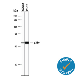
- Detection of Mouse and Rat p38 gamma by Simple WesternTM. Simple Western lane view shows lysates of C2C12 mouse myoblast cell line and PC-12 rat adrenal pheochromocytoma cell line, loaded at 0.2 mg/mL. A specific band was detected for p38 gamma at approximately 48 kDa (as indicated) using 2 µg/mL of Rabbit Anti-Human/Mouse/Rat p38 gamma Antigen Affinity-purified Polyclonal Antibody (Catalog # AF1347). This experiment was conducted under reducing conditions and using the 12-230 kDa separation system.

- Submitted by
- Novus Biologicals (provider)
- Main image
- Experimental details
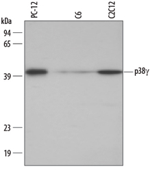
- Detection of Mouse/Rat p38 gamma by Western Blot. Western blot shows lysates of PC-12 rat adrenal pheochromocytoma cell line, C6 rat glioma cell line, and C2C12 mouse myoblast cell line. PVDF membrane was probed with 0.2 µg/mL Rabbit Anti-Human/Mouse/Rat p38 gamma Antigen Affinity-purified Polyclonal Antibody (Catalog # AF1347) followed by HRP-conjugated Anti-Rabbit IgG Secondary Antibody (Catalog # HAF008). A specific band for p38 gamma was detected at approximately 40 kDa (as indicated). This experiment was conducted under reducing conditions and using Immunoblot Buffer Group 1.
- Submitted by
- Novus Biologicals (provider)
- Main image
- Experimental details
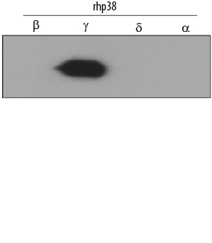
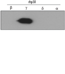
- Detection of human p38 gamma by Western Blot. Western blot shows recombinant human p38 beta , p38 gamma , p38 delta and p38 alpha (1 ng/lane). PVDF membrane was probed with 0.2 µg/mL Rabbit Anti-Human/Mouse/Rat p38 gamma Antigen Affinity-purified Polyclonal Antibody (Catalog # AF1347) followed by HRP-conjugated Anti-Rabbit IgG Secondary Antibody (Catalog # HAF008). A specific band for p38 gamma was detected at approx-imately 40 kDa (as indicated). This experiment was conducted under reducing conditions and using Immunoblot Buffer Group 1.