 Explore
 Validate
 Learn
Learn730018
antibody from Invitrogen Antibodies
Targeting: CBX3
HP1Hs-gamma
 Western blot
 Immunocytochemistry Immunoprecipitation Flow cytometry Chromatin Immunoprecipitation Other assay
Antibody data
- Antibody Data
- Antigen structure
- References [0]
- Comments [0]
- Validations
- Immunocytochemistry [2]
- Flow cytometry [1]
- Chromatin Immunoprecipitation [1]
- Other assay [1]
Submit
Validation data
Reference
Comment
Report error
- Product number
- 730018 - Provider product page

- Provider
- Invitrogen Antibodies
- Product name
- Anti-HP1 gamma Monoclonal Antibody
- Antibody type
- Monoclonal
- Antigen
- Other
- Description
- 730019 has successfully been used to detect CBX5 in mouse, human and rat. 730019 has been successfully used in western blotting and immunofluorescence to detect CBX5.
- Reactivity
- Human, Mouse, Rat
- Host
- Mouse
- Isotype
- IgG
- Vial size
- 100 µg
- Concentration
- 0.5 mg/ml
- Storage
- Maintain refrigerated at 2-8°C for up to 1 month. For long term storage store at -20°C
No comments: Submit comment
Supportive validation
- Submitted by
- Invitrogen Antibodies (provider)
- Main image
- Experimental details
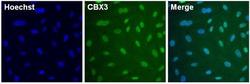
- Immunofluorescent analysis of CBX3 (green) in HeLa cells. The cells were fixed with formalin for 15 minutes, permeabilized with 1X Permeablilization buffer (Product # 8408400) for 15 minutes, and blocked with 1% Blocker BSA (Product # 37525) for 15 minutes at room temperature. Cells were stained with CBX3 monoclonal antibody (Product # 730018) at a dilution of 1:250 in blocking buffer for one hour at room temperature, washed with 1X TBS Tween 20 Buffer (Product # 28360), followed by incubation with DyLight 488 goat anti-mouse IgG secondary antibody (green, Product # A28175) at a dilution of 1:2000 and Hoechst 33342 dye (blue, Product # 62249) at a dilution of 1:5000 for 30 minutes at room temperature. Images were taken on a Thermo Scientific ToxInsight at 20X magnification.

- Submitted by
- Invitrogen Antibodies (provider)
- Main image
- Experimental details
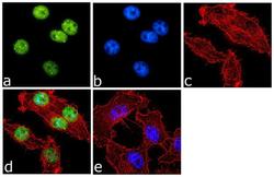
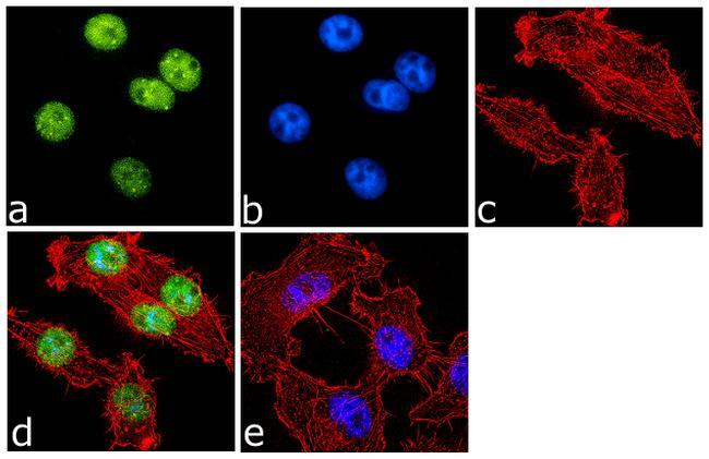
- Immunofluorescence analysis of CBX3 was done on 70% confluent log phase PC-3 cells. The cells were fixed with 4% paraformaldehyde for 10 minutes, permeabilized with 0.1% Triton™ X-100 for 10 minutes, and blocked with 1% BSA for 1 hour at room temperature. The cells were labeled with CBX3 Mouse Monoclonal Antibody (Product # 730018) at 2 µg/mL in 0.1% BSA and incubated for 3 hours at room temperature and then labeled with Goat anti-Mouse IgG (H+L) Superclonal™ Secondary Antibody, Alexa Fluor® 488 conjugate (Product # A28175) at a dilution of 1:2000 for 45 minutes at room temperature (Panel a: green). Nuclei (Panel b: blue) were stained with SlowFade® Gold Antifade Mountant with DAPI (Product # S36938). F-actin (Panel c: red) was stained with Alexa Fluor® 555 Rhodamine Phalloidin (Product # R415, 1:300). Panel d is a merged image showing nuclear localization. Panel e is a no primary antibody control. The images were captured at 60X magnification.
Supportive validation
- Submitted by
- Invitrogen Antibodies (provider)
- Main image
- Experimental details
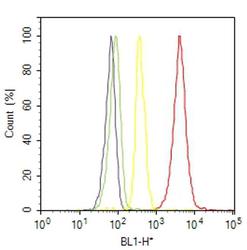
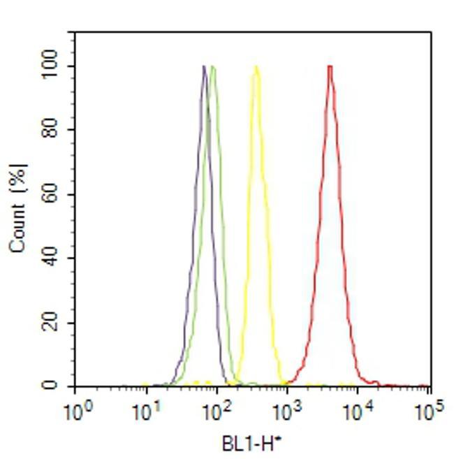
- Flow cytometry analysis of CBX3 was done on HT-29 cells. Cells were fixed with 70% ethanol for 10 minutes, permeabilized with 0.25% Triton™ X-100 for 20 minutes, and blocked with 5% BSA for 30 minutes at room temperature. Cells were labeled with CBX3 Mouse Monoclonal Antibody (730018, red histogram) or with mouse isotype control (yellow histogram) at 3-5 ug/million cells in 2.5% BSA. After incubation at room temperature for 2 hours, the cells were labeled with Alexa Fluor® 488 Rabbit Anti-Mouse Secondary Antibody (A11059) at a dilution of 1:400 for 30 minutes at room temperature. The representative 10,000 cells were acquired and analyzed for each sample using an Attune® Acoustic Focusing Cytometer. The purple histogram represents unstained control cells and the green histogram represents no-primary-antibody control.
Supportive validation
- Submitted by
- Invitrogen Antibodies (provider)
- Main image
- Experimental details
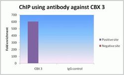
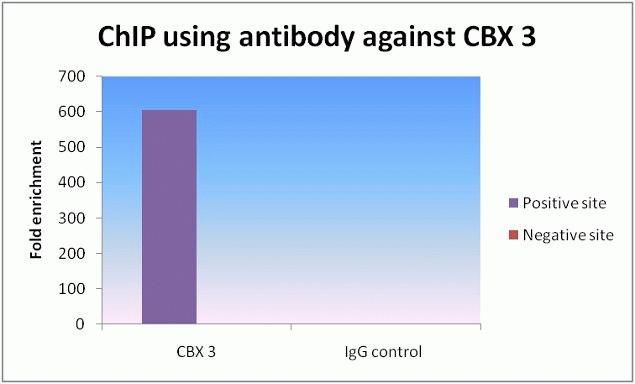
- ChIP qPCR analysis of CBX 3 Monoclonal Antibody, Recombinant (Product # 730018). ChIP was performed on HeLa chromatin using CBX 3 Mouse Monoclonal Antibody. Realtime PCR was performed for MYT and negative control locus targets using Isotype antibody as a negative control. Data is presented as fold enrichment of the ChIP antibody signal versus the negative control IgG using the comparative CT method.
Supportive validation
- Submitted by
- Invitrogen Antibodies (provider)
- Main image
- Experimental details
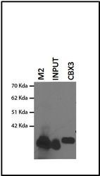
- Immunoprecipitation analysis of Anti-CBX3 Recombinant Mouse Monoclonal Antibody (Product # 730018). Immunoprecipitation analysis was performed on HEK293 cells over expressing flag tagged CBX3 protein. Western blot using anti-flag antibody was performed on the raw lysate (lane 1), and samples immunoprecipitated with Anti-CBX3 Recombinant Mouse Monoclonal Antibody (lane 2 and 3).
