 Explore
 Validate
 Learn
Learn Western blot
 ELISA
ELISA Immunocytochemistry
Antibody data
- Antibody Data
- Antigen structure
- References [0]
- Comments [0]
- Validations
- Western blot [2]
- Immunocytochemistry [5]
- Immunoprecipitation [2]
Submit
Validation data
Reference
Comment
Report error
- Product number
- LS-C812998 - Provider product page

- Provider
- LSBio
- Product name
- CBX3 / HP1 Gamma Antibody LS-C812998
- Antibody type
- Monoclonal
- Description
- Immunogen affinity purified
- Reactivity
- Human, Mouse, Rat, Hamster, Simian
- Host
- Mouse
- Isotype
- IgG
- Storage
- Store at -20°C. Avoid freeze-thaw cycles.
No comments: Submit comment
Enhanced validation
- Submitted by
- LSBio (provider)
- Enhanced method
- Genetic validation
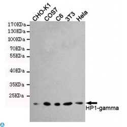
- Main image
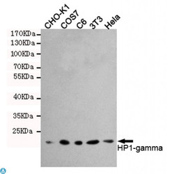
- Experimental details
- Western blot detection of HP1-gamma in Hela, 3T3, C6, COS7 and CHO-K1 cell lysates using HP1-gamma mouse mAb (1:1000 diluted). Predicted band size: 22KDa. Observed band size: 22KDa.
- Submitted by
- LSBio (provider)
- Enhanced method
- Genetic validation
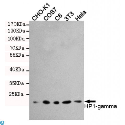
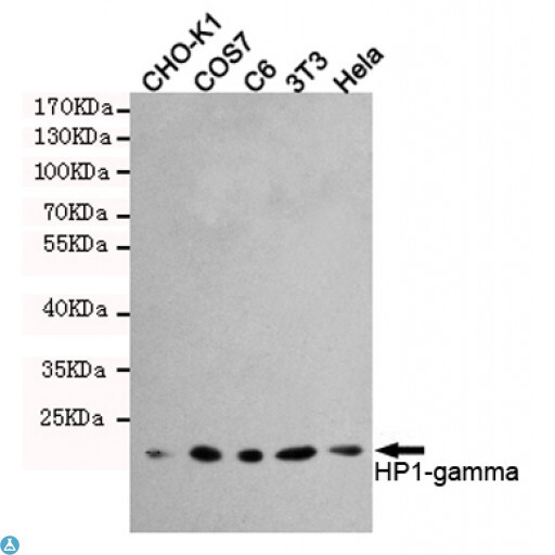
- Main image
- Experimental details
- Western blot detection of HP1-gamma in Hela, 3T3, C6, COS7 and CHO-K1 cell lysates using HP1-gamma mouse mAb (1:1000 diluted). Predicted band size: 22KDa. Observed band size: 22KDa.
Supportive validation
- Submitted by
- LSBio (provider)
- Enhanced method
- Genetic validation
- Main image
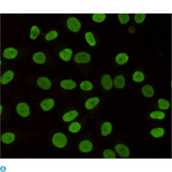
- Experimental details
- Immunocytochemistry staining of HeLa cells fixed with 4% Paraformaldehyde and using anti-HP1-gamma mouse mAb (dilution 1:200).

- Submitted by
- LSBio (provider)
- Enhanced method
- Genetic validation
- Main image

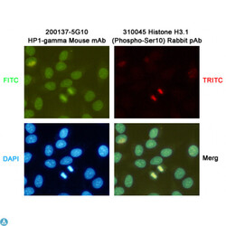
- Experimental details
- Immunocytochemistry staining of HeLa cells fixed with -20°C Methanol and using HP1-gamma (200137-5G10,dilution 1:200) mouse mAb (green) and Histone H3.1 (Phospho-Ser10) (310045,dilution 1:200) Rabbit pAb (red). DAPI was used to stain nucleus (blue).

- Submitted by
- LSBio (provider)
- Main image
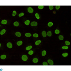
- Experimental details
- Immunocytochemistry staining of HeLa cells fixed with 4% Paraformaldehyde and using anti-HP1-gamma mouse mAb (dilution 1:200).

- Submitted by
- LSBio (provider)
- Main image

- Experimental details
- Immunocytochemistry staining of HeLa cells fixed with -20°C Methanol and using HP1-gamma (200137-5G10,dilution 1:200) mouse mAb (green) and Histone H3.1 (Phospho-Ser10) (310045,dilution 1:200) Rabbit pAb (red). DAPI was used to stain nucleus (blue).

- Submitted by
- LSBio (provider)
- Main image
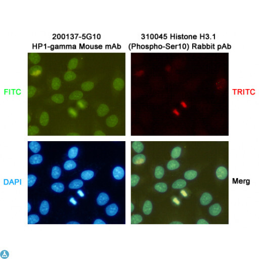
- Experimental details
- Immunocytochemistry staining of HeLa cells fixed with -20°C Methanol and using HP1-gamma (200137-5G10,dilution 1:200) mouse mAb (green) and Histone H3.1 (Phospho-Ser10) (310045,dilution 1:200) Rabbit pAb (red). DAPI was used to stain nucleus (blue).
Supportive validation
- Submitted by
- LSBio (provider)
- Enhanced method
- Genetic validation
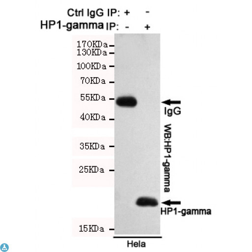
- Main image

- Experimental details
- Immunoprecipitation analysis of Hela cell lysates using HP1-gamma mouse mAb.
- Submitted by
- LSBio (provider)
- Main image
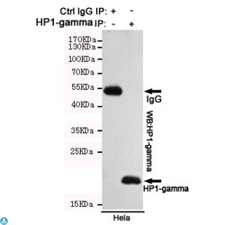
- Experimental details
- Immunoprecipitation analysis of Hela cell lysates using HP1-gamma mouse mAb.
