 Explore
 Validate
 Learn
Learn Western blot
Antibody data
- Antibody Data
- Antigen structure
- References [1]
- Comments [0]
- Validations
- Western blot [1]
- ELISA [1]
- Proximity ligation assay [1]
Submit
Validation data
Reference
Comment
Report error
- Product number
- H00000091-M09 - Provider product page

- Provider
- Abnova Corporation
- Proper citation
- Abnova Corporation Cat#H00000091-M09, RRID:AB_529955
- Product name
- ACVR1B monoclonal antibody (M09), clone 1C1
- Antibody type
- Monoclonal
- Description
- Mouse monoclonal antibody raised against a partial recombinant ACVR1B.
- Antigen sequence
SGPRGVQALLCACTSCLQANYTCETDGACMVSIFN
LDGMEHHVRTCIPKVELVPAGKPFYCLSSEDLRNT
HCCYTDYCNRIDLRVPSGHLKEPEHPSMWGPVE- Isotype
- IgG
- Antibody clone number
- 1C1
- Storage
- Store at -20°C or lower. Aliquot to avoid repeated freezing and thawing.
Submitted references Activin acutely sensitizes dorsal root ganglion neurons and induces hyperalgesia via PKC-mediated potentiation of transient receptor potential vanilloid I.
Zhu W, Xu P, Cuascut FX, Hall AK, Oxford GS
The Journal of neuroscience : the official journal of the Society for Neuroscience 2007 Dec 12;27(50):13770-80
The Journal of neuroscience : the official journal of the Society for Neuroscience 2007 Dec 12;27(50):13770-80
No comments: Submit comment
Supportive validation
- Submitted by
- Abnova Corporation (provider)
- Main image

- Experimental details
- Western Blot analysis of ACVR1B expression in transfected 293T cell line by ACVR1B monoclonal antibody (M09), clone 1C1.Lane 1: ACVR1B transfected lysate (Predicted MW: 56.8 KDa).Lane 2: Non-transfected lysate.

Supportive validation
- Submitted by
- Abnova Corporation (provider)
- Main image
- Experimental details
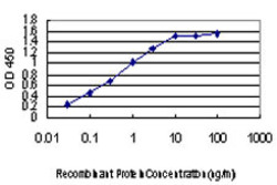
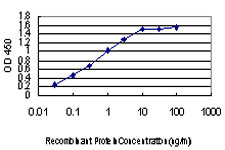
- Detection limit for recombinant GST tagged ACVR1B is approximately 0.03ng/ml as a capture antibody.
- Validation comment
- Sandwich ELISA (Recombinant protein)
- Protocol
- Protocol
Supportive validation
- Submitted by
- Abnova Corporation (provider)
- Main image

- Experimental details
- Proximity Ligation Analysis of protein-protein interactions between XIAP and ACVR1B. HeLa cells were stained with anti-XIAP rabbit purified polyclonal 1:1200 and anti-ACVR1B mouse monoclonal antibody 1:50. Each red dot represents the detection of protein-protein interaction complex, and nuclei were counterstained with DAPI (blue).
- Validation comment
- In situ Proximity Ligation Assay (Cell)
