Explore
 Validate
 Learn
Learn Western blot
 Immunocytochemistry
Antibody data
- Antibody Data
- Antigen structure
- References [0]
- Comments [0]
- Validations
- Western blot [2]
- Immunohistochemistry [1]
Submit
Validation data
Reference
Comment
Report error
- Product number
- NB100-92327 - Provider product page

- Provider
- Novus Biologicals
- Proper citation
- Novus Cat#NB100-92327, RRID:AB_1217083
- Product name
- Rabbit Polyclonal EGR2 Antibody
- Antibody type
- Polyclonal
- Description
- Immunogen affinity purified.
- Reactivity
- Human, Mouse, Rat
- Host
- Rabbit
- Isotype
- IgG
- Vial size
- 0.1 ml
- Concentration
- 1.0 mg/ml
- Storage
- Store at 4C short term. Aliquot and store at -20C long term. Avoid freeze-thaw cycles.
No comments: Submit comment
Supportive validation
- Submitted by
- Novus Biologicals (provider)
- Main image
- Experimental details
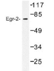
- Western Blot: EGR2 Antibody [NB100-92327] - Egr-2 Antibody in extracts from HUVEC cells.

- Submitted by
- Novus Biologicals (provider)
- Main image
- Experimental details
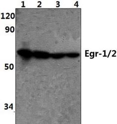
- Western Blot: EGR2 Antibody [NB100-92327] - Analysis of pAb at 1:500 dilution Lane1:HEK293T whole cell lysate(40ug) Lane2:PC12 whole cell lysate(40ug) Lane3:NIH-3T3 whole cell lysate(40ug) Lane3:Jurkat whole cell lysate(40ug)
Supportive validation
- Submitted by
- Novus Biologicals (provider)
- Main image
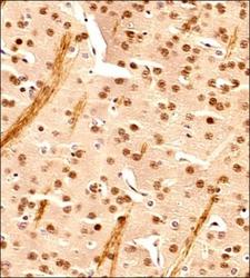
- Experimental details
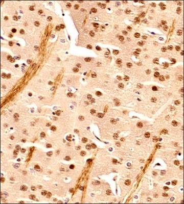
- Immunohistochemistry-Paraffin: EGR2 Antibody [NB100-92327] - IHC analysis of a formalin fixed and paraffin embedded tissue section of mouse brain using EGR2 antibody at 1:200 dilution. The signal was detected using HRP-conjugated anti-rabbit secondary antibody and DAB based detection reagent, and the sections were further counterstained with hematoxylin. This EGR2 antibody generated primarily a nuclear staining signal in the neurons and the glial cells, however, some cytoplasmic staining was also observed. The cytoplasmic signal potentially represents the translational or 14-3-3sigma interacting cytoplasmic pool of EGR2.