 Explore
 Validate
 Learn
Learn Western blot
 ELISA
ELISA Immunohistochemistry
Antibody data
- Antibody Data
- Antigen structure
- References [0]
- Comments [0]
- Validations
- Western blot [1]
- Immunocytochemistry [3]
Submit
Validation data
Reference
Comment
Report error
- Product number
- LS-B354 - Provider product page

- Provider
- LSBio
- Product name
- IHC-plus™ CDK9 Antibody (N-Terminus and C-Terminus) LS-B354
- Antibody type
- Polyclonal
- Description
- Delipidated and defibrinated
- Reactivity
- Human, Mouse, Rat
- Host
- Rabbit
- Storage
- Store vial at -20°C or below prior to opening. Dilute only prior to immediate use. Aliquot contents and freeze at -20°C or below. Avoid freeze-thaw cycles.
No comments: Submit comment
Supportive validation
- Submitted by
- LSBio (provider)
- Enhanced method
- Genetic validation
- Main image
- Experimental details
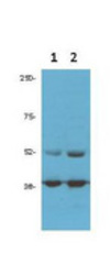
- Anti-cdk9 (PITALRE) Antibody - Western blot. Anti-cdk9 antibody (1:1500 ) was used for Western blot of 1) PC3 and 2) DU145 prostate cancer cells (50 ug per lane). Bands at the expected MW of 55 and 42 Kda were detected. Personal communication Flavio Rizzolio, Temple University.

Supportive validation
- Submitted by
- LSBio (provider)
- Enhanced method
- Genetic validation
- Main image

- Experimental details
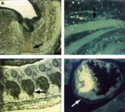
- Anti-cdk9 (PITALRE) Antibody - Immunocytochemical Staining. Immunocytochemical staining of mouse tissue using anti-cdk9 (PITALRE) antiserum. The staining shows the location of mcdk9/PITALRE protein in developing mouse tissue. Arrows indicate areas of high expression. Panel A: Peroxidase-DAB immunostaining of mcdk9/PITALRE protein in the developing mouse brain in the differentiated region of the medulla oblongata just below the fourth ventricle. Similar staining is shown in Panel B in the dorsal root ganglia. Panel C: Fluorescein immunofluorescence of mcdk9IPITALRE in skeletal muscle. Similar staining is shown in Panel D in cardiac muscle. Sections from each specimen were cut at 5-7 micron, mounted on glass and dried overnight at 37°C. All sections then were deparaffinized in xylene, rehydrated through a graded alcohol series and washed in phosphate-buffered saline (PBS). PBS was used for all subsequent washes and for antiserum dilution. Tissue sections were quenched sequentially in 0.5% hydrogen peroxide and blocked with diluted 10% normal goat anti-rabbit serum. Slides were incubated at 20° C for 1 h with rabbit anti-cdk9 (1:500) dilution, washed, and then reacted with diluted goat anti-rabbit biotinylated antibody for 30 min. All the slides were then reacted with streptavidin-peroxidase conjugate for 30 min at 20° C. Diaminobenzidine was used as the final chromogen and hematoxylin was used as the nuclear counterstain.

- Submitted by
- LSBio (provider)
- Main image
- Experimental details
- Anti-cdk9 (PITALRE) Antibody - Immunocytochemical Staining. Immunocytochemical staining of mouse tissue using anti-cdk9 (PITALRE) antiserum. The staining shows the location of mcdk9/PITALRE protein in developing mouse tissue. Arrows indicate areas of high expression. Panel A: Peroxidase-DAB immunostaining of mcdk9/PITALRE protein in the developing mouse brain in the differentiated region of the medulla oblongata just below the fourth ventricle. Similar staining is shown in Panel B in the dorsal root ganglia. Panel C: Fluorescein immunofluorescence of mcdk9IPITALRE in skeletal muscle. Similar staining is shown in Panel D in cardiac muscle. Sections from each specimen were cut at 5-7 micron, mounted on glass and dried overnight at 37°C. All sections then were deparaffinized in xylene, rehydrated through a graded alcohol series and washed in phosphate-buffered saline (PBS). PBS was used for all subsequent washes and for antiserum dilution. Tissue sections were quenched sequentially in 0.5% hydrogen peroxide and blocked with diluted 10% normal goat anti-rabbit serum. Slides were incubated at 20° C for 1 h with rabbit anti-cdk9 (1:500) dilution, washed, and then reacted with diluted goat anti-rabbit biotinylated antibody for 30 min. All the slides were then reacted with streptavidin-peroxidase conjugate for 30 min at 20° C. Diaminobenzidine was used as the final chromogen and hematoxylin was used as the nuclear counterstain.

- Submitted by
- LSBio (provider)
- Main image

- Experimental details
- Anti-cdk9 (PITALRE) Antibody - Immunocytochemical Staining. Immunocytochemical staining of mouse tissue using anti-cdk9 (PITALRE) antiserum. The staining shows the location of mcdk9/PITALRE protein in developing mouse tissue. Arrows indicate areas of high expression. Panel A: Peroxidase-DAB immunostaining of mcdk9/PITALRE protein in the developing mouse brain in the differentiated region of the medulla oblongata just below the fourth ventricle. Similar staining is shown in Panel B in the dorsal root ganglia. Panel C: Fluorescein immunofluorescence of mcdk9IPITALRE in skeletal muscle. Similar staining is shown in Panel D in cardiac muscle. Sections from each specimen were cut at 5-7 micron, mounted on glass and dried overnight at 37°C. All sections then were deparaffinized in xylene, rehydrated through a graded alcohol series and washed in phosphate-buffered saline (PBS). PBS was used for all subsequent washes and for antiserum dilution. Tissue sections were quenched sequentially in 0.5% hydrogen peroxide and blocked with diluted 10% normal goat anti-rabbit serum. Slides were incubated at 20° C for 1 h with rabbit anti-cdk9 (1:500) dilution, washed, and then reacted with diluted goat anti-rabbit biotinylated antibody for 30 min. All the slides were then reacted with streptavidin-peroxidase conjugate for 30 min at 20° C. Diaminobenzidine was used as the final chromogen and hematoxylin was used as the nuclear counterstain.
