 Explore
 Validate
 Learn
Learn Western blot
 Immunocytochemistry
Antibody data
- Antibody Data
- Antigen structure
- References [6]
- Comments [0]
- Validations
- Western blot [2]
Submit
Validation data
Reference
Comment
Report error
- Product number
- ABIN359071 - Provider product page

- Provider
- antibodies-online
- Product name
- anti-Protein Kinase D3 (PRKD3) (Middle Region) antibody
- Antibody type
- Polyclonal
- Antigen
- This antibody is generated from rabbits immunized with a KLH conjugated synthetic peptide selected from the center region of human PKC nu.
- Description
- Protein G column, eluted with high and low pH buffers and neutralized immediately, followed by dialysis against PBS
- Reactivity
- Human
- Host
- Rabbit
- Epitope
- Middle Region
- Vial size
- 0.4 mL
- Concentration
- 0.25 mg/mL
- Storage
- Store the antibody undiluted at 2-8°C for one month or (in aliquots) at-20°C for longer.
- Handling
- Avoid repeated freezing and thawing.
Submitted references Protein kinase D3 (PKD3) contributes to prostate cancer cell growth and survival through a PKCepsilon/PKD3 pathway downstream of Akt and ERK 1/2.
Protein kinase D regulates basolateral membrane protein exit from trans-Golgi network.
Regulation of protein kinase Cnu by the B-cell antigen receptor.
Protein kinase C nu/protein kinase D3 nuclear localization, catalytic activation, and intracellular redistribution in response to G protein-coupled receptor agonists.
Signaling pathways triggered by HIV-1 Tat in human monocytes to induce TNF-alpha.
HIV-1 Tat protein induces interleukin-10 in human peripheral blood monocytes: involvement of protein kinase C-betaII and -delta.
Chen J, Deng F, Singh SV, Wang QJ
Cancer research 2008 May 15;68(10):3844-53
Cancer research 2008 May 15;68(10):3844-53
Protein kinase D regulates basolateral membrane protein exit from trans-Golgi network.
Yeaman C, Ayala MI, Wright JR, Bard F, Bossard C, Ang A, Maeda Y, Seufferlein T, Mellman I, Nelson WJ, Malhotra V
Nature cell biology 2004 Feb;6(2):106-12
Nature cell biology 2004 Feb;6(2):106-12
Regulation of protein kinase Cnu by the B-cell antigen receptor.
Matthews SA, Dayalu R, Thompson LJ, Scharenberg AM
The Journal of biological chemistry 2003 Mar 14;278(11):9086-91
The Journal of biological chemistry 2003 Mar 14;278(11):9086-91
Protein kinase C nu/protein kinase D3 nuclear localization, catalytic activation, and intracellular redistribution in response to G protein-coupled receptor agonists.
Rey O, Yuan J, Young SH, Rozengurt E
The Journal of biological chemistry 2003 Jun 27;278(26):23773-85
The Journal of biological chemistry 2003 Jun 27;278(26):23773-85
Signaling pathways triggered by HIV-1 Tat in human monocytes to induce TNF-alpha.
Bennasser Y, Badou A, Tkaczuk J, Bahraoui E
Virology 2002 Nov 10;303(1):174-80
Virology 2002 Nov 10;303(1):174-80
HIV-1 Tat protein induces interleukin-10 in human peripheral blood monocytes: involvement of protein kinase C-betaII and -delta.
Bennasser Y, Bahraoui E
FASEB journal : official publication of the Federation of American Societies for Experimental Biology 2002 Apr;16(6):546-54
FASEB journal : official publication of the Federation of American Societies for Experimental Biology 2002 Apr;16(6):546-54
No comments: Submit comment
Supportive validation
- Submitted by
- antibodies-online (provider)
- Main image
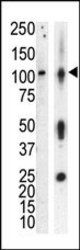
- Experimental details
- Western blot analysis of anti-PKCnu Pab in lysate of HL60 cells stimulated with PMA (lane A) and mouse brain tissue lysate (lane B). PKCnu (arrow) was detected using purified Pab. Secondary HRP-anti-rabbit was used for signal visualization with chemiluminescence.

- Submitted by
- antibodies-online (provider)
- Main image

- Experimental details
- TOP: Upper panel, western blot analysis of GFP fusion protein expression in Panc-1 cells by using an anti-GFP antibody. Lanes 1 and 5: non-transfected cells; lanes 2 and 6: GFP-PKD-transfected cells; lanes 3 and 7: GFP-PKD2-transfected cells; lanes 4 and 8: GFP-PKD3 transfected cells. Center panel, western blot analysis of GFP fusion protein expression in Panc-1 cells by using PKD3 N-term (AP13555PU-N) and C-term (AP13556PU-N) antibodies. Lower panel, indirect immunofluorescence analysis of GFP-PKD3 fusion protein expression in Panc-1 cells by using AP13555PU-N and AP13556PU-N antibodies. BOTTOM: Formalin-fixed and paraffin-embedded human cancer tissue reacted with the primary antibody, which was peroxidase-conjugated to the secondary antibody, followed by AEC staining.
