Explore
 Validate
 Learn
Learn Western blot
Antibody data
- Antibody Data
- Antigen structure
- References [1]
- Comments [0]
- Validations
- Western blot [2]
- Immunocytochemistry [1]
- Immunohistochemistry [1]
- Other assay [1]
Submit
Validation data
Reference
Comment
Report error
- Product number
- PA5-14938 - Provider product page

- Provider
- Invitrogen Antibodies
- Product name
- LIMK1 Polyclonal Antibody
- Antibody type
- Polyclonal
- Antigen
- Synthetic peptide
- Reactivity
- Human
- Host
- Rabbit
- Isotype
- IgG
- Vial size
- 400 µL
- Concentration
- 2 mg/mL
- Storage
- Store at 4°C short term. For long term storage, store at -20°C, avoiding freeze/thaw cycles.
Submitted references Cofilin-1, LIMK1 and SSH1 are differentially expressed in locally advanced colorectal cancer and according to consensus molecular subtypes.
Sousa-Squiavinato ACM, Vasconcelos RI, Gehren AS, Fernandes PV, de Oliveira IM, Boroni M, Morgado-Díaz JA
Cancer cell international 2021 Jan 22;21(1):69
Cancer cell international 2021 Jan 22;21(1):69
No comments: Submit comment
Supportive validation
- Submitted by
- Invitrogen Antibodies (provider)
- Main image

- Experimental details
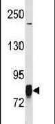
- Western blot analysis using a LIM Kinase 1 polyclonal antibody (Product # PA5-14938) in SK-BR-3 cell lysates (35 µg per lane).
- Submitted by
- Invitrogen Antibodies (provider)
- Main image
- Experimental details
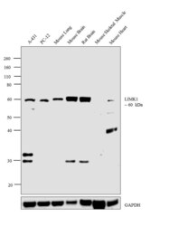
- Western blot analysis was performed on whole cell extracts (30 µg lysate) of A-431 (Lane 1), PC-12 (Lane 2) and tissue extracts (30 µg lysate) of Mouse Lung (Lane 3), Mouse Brain (Lane 4), Mouse Skeletal Muscle (Lane 5) and Mouse Heart (Lane 6). The blot was probed with Anti-LIMK1 Polyclonal Antibody (Product # PA5-14938, 1:1000 dilution) and detected by chemiluminescence using Goat anti-Rabbit IgG (H+L) Superclonal™ Secondary Antibody, HRP conjugate (Product # A27036, 0.25 µg/ml, 1:4000 dilution). A 60 kDa band corresponding to LIMK1 was not detected in Mouse Skeletal Muscle and Mouse Heart which are reported negative for LIMK1.

Supportive validation
- Submitted by
- Invitrogen Antibodies (provider)
- Main image
- Experimental details
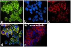
- Immunofluorescence analysis of LIMK1 was performed using 70% confluent log phase PC-12 cells. The cells were fixed with 4% paraformaldehyde for 10 minutes, permeabilized with 0.1% Triton™ X-100 for 15 minutes, and blocked with 1% BSA for 1 hour at room temperature. The cells were labeled with LIMK1 Polyclonal Antibody (Product # PA5-14938) at 1:250 dilution in 0.1% BSA, incubated at 4 degree Celsius overnight and then labeled with Goat anti-Rabbit IgG (H+L) Superclonal™ Secondary Antibody, Alexa Fluor® 488 conjugate (Product # A27034) at a dilution of 1:2000 for 45 minutes at room temperature (Panel a: green). Nuclei (Panel b: blue) were stained with ProLong™ Diamond Antifade Mountant with DAPI (Product # P36962). F-actin (Panel c: red) was stained with Rhodamine Phalloidin (Product # R415). Panel d represents the merged image showing cytoplasmic localization. Panel e represents control cells with no primary antibody to assess background. The images were captured at 60X magnification.

Supportive validation
- Submitted by
- Invitrogen Antibodies (provider)
- Main image

- Experimental details
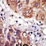
- Immunohistochemical analysis of formalin-fixed, paraffin-embedded human cancer tissue using a LIM Kinase 1 polyclonal antibody (Product # PA5-14938), followed by HRP-conjugated secondary antibody and DAB staining.
Supportive validation
- Submitted by
- Invitrogen Antibodies (provider)
- Main image
- Experimental details
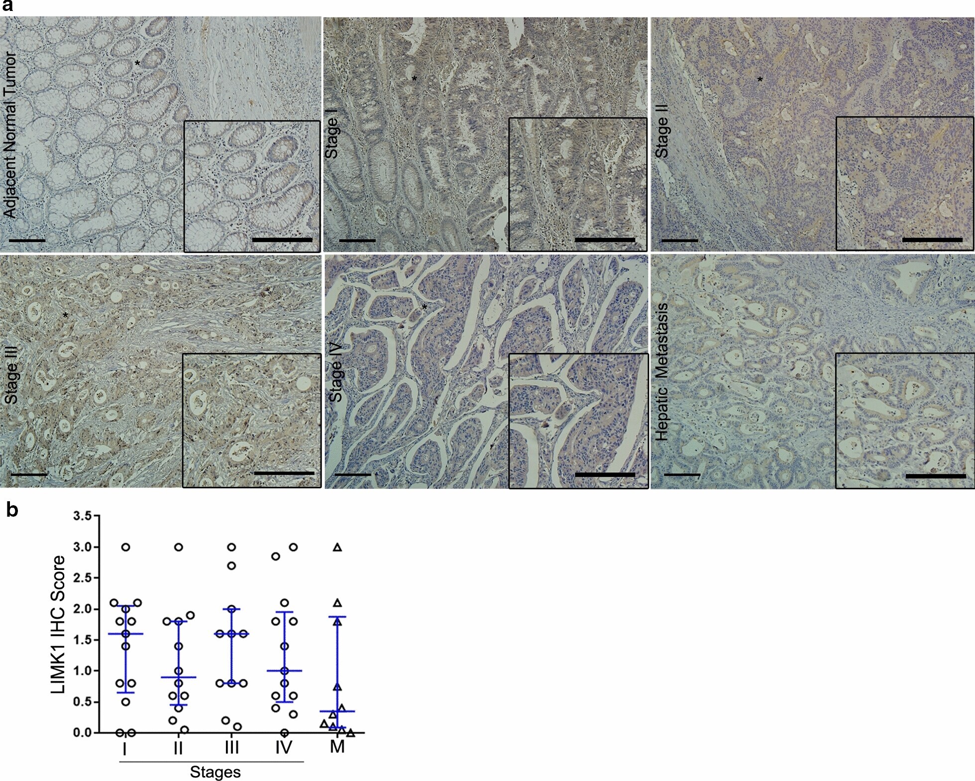
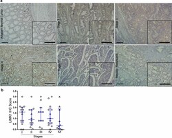
- Fig. 5 Analysis of LIMK1 protein levels in CRC tissues using IHC. a Representative IHC images for LIMK1 staining in adjacent normal tissues, tumor stages (I-IV), and hepatic metastatic lesions. Scale bar = 100 um. b Quantification of the IHC score data; this value was calculated using the intensity x area of staining. IHC and immunohistochemistry of 49 primary tumors (stages I, n = 13; II, n = 12; III, n = 11; and IV, n = 13), and 10 hepatic metastases