Explore
 Validate
 Learn
Learn ELISA
ELISA Other assay
Antibody data
- Antibody Data
- Antigen structure
- References [3]
- Comments [0]
- Validations
- Other assay [1]
Submit
Validation data
Reference
Comment
Report error
- Product number
- MA1-18418 - Provider product page

- Provider
- Invitrogen Antibodies
- Product name
- NGFR Monoclonal Antibody (ME20.4)
- Antibody type
- Monoclonal
- Antigen
- Other
- Description
- Reconstitute with the addition of 100 µL of sterile water. Centrifuge to remove any insoluble material. After reconstitution keep aliquots at -20 °C for a higher stability, and at 4 °C with an appropriate antibacterial agent. Glycerol (1:1) may be added for an additional stability. Avoid repetitive freeze/thaw cycles.
- Reactivity
- Human, Canine, Feline, Porcine, Rabbit
- Host
- Mouse
- Isotype
- IgG
- Antibody clone number
- ME20.4
- Vial size
- 100 µg
- Concentration
- 1 mg/mL
- Storage
- -20° C, Avoid Freeze/Thaw Cycles
Submitted references A single-cell atlas of the airway epithelium reveals the CFTR-rich pulmonary ionocyte.
Functional sphere profiling reveals the complexity of neuroblastoma tumor-initiating cell model.
Functional sphere profiling reveals the complexity of neuroblastoma tumor-initiating cell model.
Plasschaert LW, Žilionis R, Choo-Wing R, Savova V, Knehr J, Roma G, Klein AM, Jaffe AB
Nature 2018 Aug;560(7718):377-381
Nature 2018 Aug;560(7718):377-381
Functional sphere profiling reveals the complexity of neuroblastoma tumor-initiating cell model.
Coulon A, Flahaut M, Mühlethaler-Mottet A, Meier R, Liberman J, Balmas-Bourloud K, Nardou K, Yan P, Tercier S, Joseph JM, Sommer L, Gross N
Neoplasia (New York, N.Y.) 2011 Oct;13(10):991-1004
Neoplasia (New York, N.Y.) 2011 Oct;13(10):991-1004
No comments: Submit comment
Supportive validation
- Submitted by
- Invitrogen Antibodies (provider)
- Main image
- Experimental details
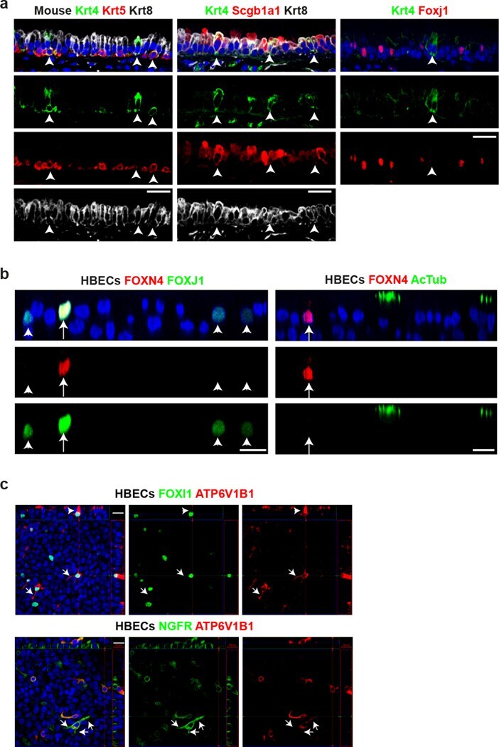
- Extended Data Figure 4: Validation of novel lineages in mouse and human by immunofluorescence. a , Immunofluorescence in mouse tracheal epithelium for Krt4 (green, arrowheads), Krt5 (basal), Krt8 (luminal), Scgb1a1 (club, secretory) and Foxj1 (ciliated) (n=3 animals). b , Immunofluorescence in differentiated HBEC cultures for FOXN4 (red, arrows), FOXJ1 (arrowheads mark FOXJ1 low cells) and Acetylated alphaTubulin (cilia) (n=2 donors). c , Immunofluorescence in HBEC cultures for the ionocyte markers FOXI1, ATP6V1B1 and NGFR (n=3 donors). Arrowhead shows apical enrichment of ATP6V1B1. Arrows highlight lateral protrusions. Scale bar, 20 mum.