Explore
 Validate
 Learn
Learn Western blot
Antibody data
- Antibody Data
- Antigen structure
- References [6]
- Comments [0]
- Validations
- Western blot [1]
- Immunohistochemistry [1]
Submit
Validation data
Reference
Comment
Report error
- Product number
- AF1157 - Provider product page

- Provider
- R&D Systems
- Product name
- Mouse NGFR/TNFRSF16 Antibody
- Antibody type
- Polyclonal
- Description
- Immunogen affinity purified. Detects mouse NGF R/TNFRSF16 in direct ELISAs and Western blots. In direct ELISAs and Western blots, approximately 5% cross-reactivity with recombinant human NGF R/TNFRSF16 is observed.
- Reactivity
- Mouse
- Host
- Goat
- Conjugate
- Unconjugated
- Antigen sequence
Q9Z0W1- Isotype
- IgG
- Vial size
- 100 ug
- Concentration
- LYOPH
- Storage
- Use a manual defrost freezer and avoid repeated freeze-thaw cycles. 12 months from date of receipt, -20 to -70 °C as supplied. 1 month, 2 to 8 °C under sterile conditions after reconstitution. 6 months, -20 to -70 °C under sterile conditions after reconstitution.
Submitted references A Novel Neuroprotective Mechanism for Lithium That Prevents Association of the p75NTR-Sortilin Receptor Complex and Attenuates proNGF-Induced Neuronal Death In Vitro and In Vivo.
Urinary Extracellular Domain of Neurotrophin Receptor p75 as a Biomarker for Amyotrophic Lateral Sclerosis in a Chinese cohort.
Superior Cervical Ganglia Neurons Induce Foxp3+ Regulatory T Cells via Calcitonin Gene-Related Peptide.
An intracellular domain fragment of the p75 neurotrophin receptor (p75(NTR)) enhances tropomyosin receptor kinase A (TrkA) receptor function.
Long-term delivery of nerve growth factor by encapsulated cell biodelivery in the Göttingen minipig basal forebrain.
Mouse natural killer (NK) cells express the nerve growth factor receptor TrkA, which is dynamically regulated.
Greenwood SG, Montroull L, Volosin M, Scharfman HE, Teng KK, Light M, Torkin R, Maxfield F, Hempstead BL, Friedman WJ
eNeuro 2018 Jan-Feb;5(1)
eNeuro 2018 Jan-Feb;5(1)
Urinary Extracellular Domain of Neurotrophin Receptor p75 as a Biomarker for Amyotrophic Lateral Sclerosis in a Chinese cohort.
Jia R, Shepheard S, Jin J, Hu F, Zhao X, Xue L, Xiang L, Qi H, Qu Q, Guo F, Rogers ML, Dang J
Scientific reports 2017 Jul 11;7(1):5127
Scientific reports 2017 Jul 11;7(1):5127
Superior Cervical Ganglia Neurons Induce Foxp3+ Regulatory T Cells via Calcitonin Gene-Related Peptide.
Szklany K, Ruiter E, Mian F, Kunze W, Bienenstock J, Forsythe P, Karimi K
PloS one 2016;11(3):e0152443
PloS one 2016;11(3):e0152443
An intracellular domain fragment of the p75 neurotrophin receptor (p75(NTR)) enhances tropomyosin receptor kinase A (TrkA) receptor function.
Matusica D, Skeldal S, Sykes AM, Palstra N, Sharma A, Coulson EJ
The Journal of biological chemistry 2013 Apr 19;288(16):11144-54
The Journal of biological chemistry 2013 Apr 19;288(16):11144-54
Long-term delivery of nerve growth factor by encapsulated cell biodelivery in the Göttingen minipig basal forebrain.
Fjord-Larsen L, Kusk P, Tornøe J, Juliusson B, Torp M, Bjarkam CR, Nielsen MS, Handberg A, Sørensen JC, Wahlberg LU
Molecular therapy : the journal of the American Society of Gene Therapy 2010 Dec;18(12):2164-72
Molecular therapy : the journal of the American Society of Gene Therapy 2010 Dec;18(12):2164-72
Mouse natural killer (NK) cells express the nerve growth factor receptor TrkA, which is dynamically regulated.
Ralainirina N, Brons NH, Ammerlaan W, Hoffmann C, Hentges F, Zimmer J
PloS one 2010 Dec 1;5(12):e15053
PloS one 2010 Dec 1;5(12):e15053
No comments: Submit comment
Supportive validation
- Submitted by
- R&D Systems (provider)
- Main image

- Experimental details
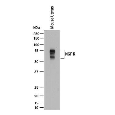
- Detection of Mouse NGF R/TNFRSF16 by Western Blot. Western blot shows lysates of mouse uterus tissue. PVDF membrane was probed with 2 µg/mL of Goat Anti-Mouse NGF R/TNFRSF16 Antigen Affinity-purified Polyclonal Antibody (Catalog # AF1157) followed by HRP-conjugated Anti-Goat IgG Secondary Antibody (Catalog # HAF017). Specific bands were detected for NGF R/TNFRSF16 at approximately 60-75 kDa (as indicated). This experiment was conducted under reducing conditions and using Immunoblot Buffer Group 1.
Supportive validation
- Submitted by
- R&D Systems (provider)
- Main image

- Experimental details
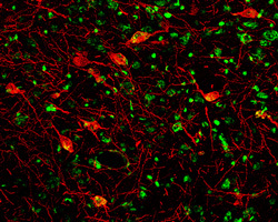
- NGF R/TNFRSF16 in Mouse Brain. NGF R/TNFRSF16 was detected in perfusion fixed frozen sections of mouse brain (cortex) using 7 µg/mL Goat Anti-Mouse NGF R/TNFRSF16 Antigen Affinity-purified Polyclonal Antibody (Catalog # AF1157) overnight at 4 °C. Tissue was stained (red) and counterstained (green). View our protocol for Fluorescent IHC Staining of Frozen Tissue Sections.