 Explore
 Validate
 Learn
Learn Western blot
Antibody data
- Antibody Data
- Antigen structure
- References [5]
- Comments [0]
- Validations
- Western blot [1]
- Immunocytochemistry [2]
- Immunohistochemistry [1]
- Flow cytometry [1]
Submit
Validation data
Reference
Comment
Report error
- Product number
- MAB367 - Provider product page

- Provider
- R&D Systems
- Product name
- Human/Canine NGFR/TNFRSF16 Antibody
- Antibody type
- Monoclonal
- Description
- Protein A or G purified from hybridoma culture supernatant. Detects human NGF R in direct ELISAs and Western blots. In direct ELISAs, no crossreactivity with recombinant human (rh) 4-1BB, rhCD27, rhCD40, rhBAFF R, rhCD30, rhDR3, rhDR6, rhEDAR, rhFas, rhHVEM, rhGITR, rhLTR B, recominant mouse (rm) NGF R, rhOPG, rmOX40, rhRANK, rhTAJ, rhTNF RI or rhTNF RII is observed.
- Reactivity
- Human, Canine
- Host
- Mouse
- Conjugate
- Unconjugated
- Antigen sequence
P08138- Isotype
- IgG
- Antibody clone number
- 74902
- Vial size
- 100 ug
- Concentration
- LYOPH
- Storage
- Use a manual defrost freezer and avoid repeated freeze-thaw cycles. 12 months from date of receipt, -20 to -70 °C as supplied. 1 month, 2 to 8 °C under sterile conditions after reconstitution. 6 months, -20 to -70 °C under sterile conditions after reconstitution.
Submitted references An initial investigation into endothelial CC chemokine expression in the human rheumatoid synovium.
Decreased demand for olfactory periglomerular cells impacts on neural precursor cell viability in the rostral migratory stream.
Human papillomavirus 16 infection predicts poor outcome in patients with esophageal squamous cell carcinoma.
BDNF and its receptors in human myasthenic thymus: implications for cell fate in thymic pathology.
Mesenchymal stem cell abnormalities in patients with multiple myeloma.
Rump L, Mattey DL, Kehoe O, Middleton J
Cytokine 2017 Sep;97:133-140
Cytokine 2017 Sep;97:133-140
Decreased demand for olfactory periglomerular cells impacts on neural precursor cell viability in the rostral migratory stream.
Langenfurth A, Gu S, Bautze V, Zhang C, Neumann JE, Schüller U, Stock K, Wolf SA, Maier AM, Mastrella G, Pak A, Cheng H, Kälin RE, Holmbeck K, Strotmann J, Kettenmann H, Glass R
Scientific reports 2016 Aug 30;6:32203
Scientific reports 2016 Aug 30;6:32203
Human papillomavirus 16 infection predicts poor outcome in patients with esophageal squamous cell carcinoma.
Xi R, Zhang X, Chen X, Pan S, Hui B, Zhang L, Fu S, Li X, Zhang X, Gong T, Guo J, Che S
OncoTargets and therapy 2015;8:573-81
OncoTargets and therapy 2015;8:573-81
BDNF and its receptors in human myasthenic thymus: implications for cell fate in thymic pathology.
Berzi A, Ayata CK, Cavalcante P, Falcone C, Candiago E, Motta T, Bernasconi P, Hohlfeld R, Mantegazza R, Meinl E, Farina C
Journal of neuroimmunology 2008 Jul 15;197(2):128-39
Journal of neuroimmunology 2008 Jul 15;197(2):128-39
Mesenchymal stem cell abnormalities in patients with multiple myeloma.
Garderet L, Mazurier C, Chapel A, Ernou I, Boutin L, Holy X, Gorin NC, Lopez M, Doucet C, Lataillade JJ
Leukemia & lymphoma 2007 Oct;48(10):2032-41
Leukemia & lymphoma 2007 Oct;48(10):2032-41
No comments: Submit comment
Supportive validation
- Submitted by
- R&D Systems (provider)
- Main image

- Experimental details
- Detection of Human NGF R/TNFRSF16 by Western Blot. Western blot shows lysates of SW480 human colorectal adenocarcinoma cell line. PVDF membrane was probed with 2 µg/mL of Mouse Anti-Human/Canine NGF R/TNFRSF16 Monoclonal Antibody (Catalog # MAB367) followed by HRP-conjugated Anti-Mouse IgG Secondary Antibody (Catalog # HAF018). A specific band was detected for NGF R/TNFRSF16 at approximately 65 kDa (as indicated). GAPDH (Catalog # MAB5718) is shown as a loading control. This experiment was conducted under non-reducing conditions and using Immunoblot Buffer Group 1.

Supportive validation
- Submitted by
- R&D Systems (provider)
- Main image

- Experimental details
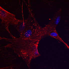
- NGF R/TNFRSF16 in Canine Mesenchymal Stem Cells. NGF R/TNFRSF16 was detected in immersion fixed canine mesenchymal stem cells using Mouse Anti-Human/Canine NGF R/TNFRSF16 Monoclonal Antibody (Catalog # MAB367) at 10 µg/mL for 3 hours at room temperature. Cells were stained using the NorthernLights™ 557-conjugated Anti-Goat IgG Secondary Antibody (red; Catalog # NL001) and counterstained with DAPI (blue). Specific staining was localized to cell surfaces. View our protocol for Fluorescent ICC Staining of Stem Cells on Coverslips.
- Submitted by
- R&D Systems (provider)
- Main image

- Experimental details
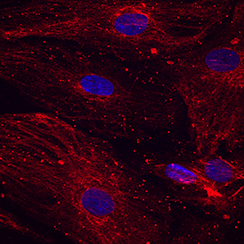
- NGF R/TNFRSF16 in Human Mesenchymal Stem Cells. NGF R/TNFRSF16 was detected in immersion fixed human mesenchymal stem cells using Mouse Anti-Human/Canine NGF R/TNFRSF16 Monoclonal Antibody (Catalog # MAB367) at 10 µg/mL for 3 hours at room temperature. Cells were stained using the NorthernLights™ 557-conjugated Anti-Goat IgG Secondary Antibody (red; Catalog # NL001) and counterstained with DAPI (blue). Specific staining was localized to cell surfaces. View our protocol for Fluorescent ICC Staining of Stem Cells on Coverslips.
Supportive validation
- Submitted by
- R&D Systems (provider)
- Main image
- Experimental details
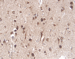
- NGF R/TNFRSF16 in Human Brain. NGF R/TNFRSF16 was detected in immersion fixed paraffin-embedded sections of human brain (cortex) using 25 µg/mL Mouse Anti-Human/Canine NGF R/TNFRSF16 Monoclonal Antibody (Catalog # MAB367) overnight at 4 °C. Tissue was stained with the Anti-Mouse HRP-DAB Cell & Tissue Staining Kit (brown; Catalog # CTS002) and counterstained with hematoxylin (blue). View our protocol for Chromogenic IHC Staining of Paraffin-embedded Tissue Sections.

Supportive validation
- Submitted by
- R&D Systems (provider)
- Main image
- Experimental details
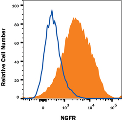
- Detection of NGF R/TNFRSF16 in SH-SY5Y Human Cell Line by Flow Cytometry. SH-SY5Y human neuroblastoma cell line was stained with Mouse Anti-Human NGF R/TNFRSF16 Monoclonal Antibody (Catalog # MAB367, filled histogram) or isotype control antibody (Catalog # MAB002, open histogram) followed by anti-Mouse IgG PE-conjugated Secondary Antibody (Catalog # F0102B). View our protocol for Staining Membrane-associated Proteins.
