Explore
 Validate
 Learn
Learn Western blot
Antibody data
- Antibody Data
- Antigen structure
- References [0]
- Comments [0]
- Validations
- Western blot [1]
- Immunocytochemistry [2]
Submit
Validation data
Reference
Comment
Report error
- Product number
- TA319603 - Provider product page

- Provider
- OriGene
- Product name
- Mouse monoclonal Keratin antibody
- Antibody type
- Monoclonal
- Description
- Mouse monoclonal Keratin antibody
- Host
- Mouse
- Conjugate
- Unconjugated
- Epitope
- KRT6C
- Antibody clone number
- C11
- Vial size
- 100 µg
- Concentration
- NULL
No comments: Submit comment
Supportive validation
- Submitted by
- OriGene (provider)
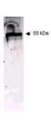
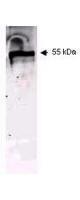
- Main image
- Experimental details
- WB using Immunochemical's Mouse Anti-Keratin antibody. This antibody recognizes a single 56 kDa band corresponding to human keratin as confirmed by the position of molecular weight markers (not shown). A 1:400 dilution of Mab anti-Keratin was used for 2h followed by detection using a 1:5,000 dilution of IRDyeTM800 conjugated Goat-a-Mouse IgG [H&L] and visualization using the Odyssey? Infrared Imaging System developed by LI-COR.
- Validation comment
- WB
Supportive validation
- Submitted by
- OriGene (provider)
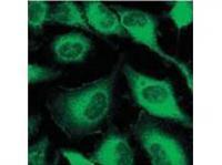
- Main image

- Experimental details
- Immunofluorescence using Immunochemical's Mouse Anti-Keratin antibody. Confocal slices of HeLa cells are between 0.5 and 0.6 ?m where the image is taken near the bottom of the cell. Use FITC a 1:2,000 dilution of FITC conjugated Goat-a-Mouse IgG [H&L] (610-102-121) for detection
- Validation comment
- IF
- Submitted by
- OriGene (provider)
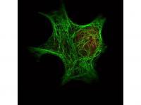
- Main image

- Experimental details
- IF of Immunochemical's Anti-Keratin antibody (TA319603) was used with Dylight 488 goat anti-mouse (shown in green) to detect Keratin by IF. In the same experiment, polyclonal Anti-HDAC-1 antibody (TA319251) was used with Atto425 Anti-Rabbit IgG (shown in red) to detect HDAC-1.
- Validation comment
- IF