Explore
 Validate
 Learn
Learn Western blot
Antibody data
- Antibody Data
- Antigen structure
- References [0]
- Comments [0]
- Validations
- Western blot [6]
- Immunocytochemistry [1]
- Immunohistochemistry [2]
- Chromatin Immunoprecipitation [1]
Submit
Validation data
Reference
Comment
Report error
- Product number
- PA5-78191 - Provider product page

- Provider
- Invitrogen Antibodies
- Product name
- TCF7 Polyclonal Antibody
- Antibody type
- Polyclonal
- Antigen
- Recombinant full-length protein
- Description
- Positive Control: HepG2, Jurkat
- Concentration
- 1 mg/mL
No comments: Submit comment
Supportive validation
- Submitted by
- Invitrogen Antibodies (provider)
- Main image

- Experimental details
- Western blot analysis of TCF7 in Jurkat whole cell lysate using 30 µg of protein. Samples were separated with 10% SDS-PAGE and incubated with TCF7 polyclonal antibody (Product # PA5-78191) using a dilution of 1:1000.

- Submitted by
- Invitrogen Antibodies (provider)
- Main image

- Experimental details
- Western blot analysis of TCF7 in HepG2 cells using 30 µg of protein. Samples were separated with 10% SDS-PAGE and incubated with TCF7 polyclonal antibody (Product # PA5-78191) using a dilution of 1:1000.
- Submitted by
- Invitrogen Antibodies (provider)
- Main image
- Experimental details
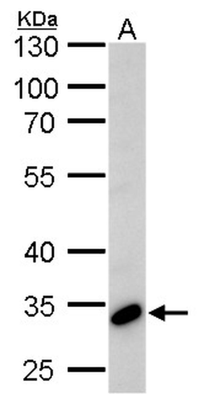
- TCF7 Polyclonal Antibody detects TCF7 protein by Western blot analysis. A. 30 µg Jurkat whole cell lysate/extract.10 % SDS-PAGE. TCF7 Polyclonal Antibody (Product # PA5-78191) dilution: 1:1,000.
- Submitted by
- Invitrogen Antibodies (provider)
- Main image
- Experimental details
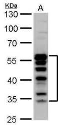
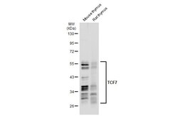
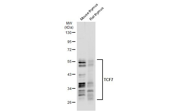
- Western Blot using TCF7 Polyclonal Antibody (Product # PA5-78191). Various tissue extracts (50 µg) were separated by 10% SDS-PAGE, and the membrane was blotted with TCF7 Polyclonal Antibody (Product # PA5-78191) diluted at 1:500. The HRP-conjugated anti-rabbit IgG antibody was used to detect the primary antibody.
- Submitted by
- Invitrogen Antibodies (provider)
- Main image

- Experimental details
- TCF7 Polyclonal Antibody detects TCF7 protein by Western blot analysis. A. 30 µg Jurkat whole cell lysate/extract.10 % SDS-PAGE. TCF7 Polyclonal Antibody (Product # PA5-78191) dilution: 1:1,000.

- Submitted by
- Invitrogen Antibodies (provider)
- Main image
- Experimental details
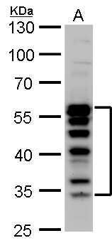
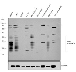
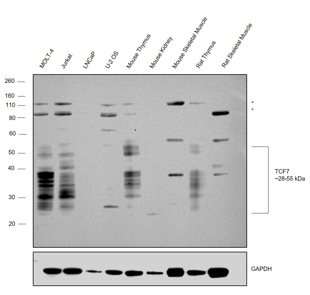
- Western blot was performed using Anti-TCF7 Polyclonal Antibody (Product # PA5-78191) and bands corresponding to TCF7 at a range between 28-55 kDa was observed across the panel tested except in LNCaP, U-2 OS, Mouse kidney, Mouse and Rat Skeletal Muscle which are low expressors of TCF7. Modified whole cell extracts (1% SDS) (30 µg lysate) of MOLT-4 (Lane 1), Jurkat (Lane 2), LNCaP (Lane 3), U-2 OS (Lane 4) and tissue extracts (30 µg lysate) of Mouse Thymus (Lane 5), Mouse Kidney (Lane 6), Mouse Skeletal Muscle (Lane 7), Rat Thymus (Lane 8) and Rat Skeletal Muscle (Lane 9) were electrophoresed using NuPAGE™ 4-12% Bis-Tris Protein Gel (Product # NP0321BOX). Resolved proteins were then transferred onto a Nitrocellulose membrane (Product # IB23001) by iBlot® 2 Dry Blotting System (Product # IB21001). The blot was probed with the primary antibody (1:2000 dilution) and detected by chemiluminescence with Goat anti-Rabbit IgG (H+L) Superclonal™ Recombinant Secondary Antibody, HRP (Product # A27036, 1:4000 dilution) using the iBright FL 1000 (Product # A32752). Chemiluminescent detection was performed using Novex® ECL Reagent Kit (Product # WP20005). Few uncharacterized bands (*) were also observed across the panel.
Supportive validation
- Submitted by
- Invitrogen Antibodies (provider)
- Main image
- Experimental details
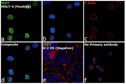
- Immunofluorescence analysis of TCF7 was performed using MOLT-4 and U-2 OS cells. The cells were fixed with 4% paraformaldehyde for 10 minutes, permeabilized with 0.1% Triton™ X-100 for 15 minutes, and blocked with 2% BSA for 1 hour at room temperature. The cells were labeled with TCF7 Polyclonal Antibody (Product # PA5-78191) at 1:200 dilution in 0.1% BSA and incubated overnight at 4 degree and then labeled with Goat anti-Rabbit IgG (H+L) Highly Cross-Adsorbed Secondary Antibody, Alexa Fluor Plus 488 (Product # A32731) at a dilution of 1:2000 for 45 minutes at room temperature (Panel a: green). Nuclei (Panel b: blue) were stained with ProLong™ Diamond Antifade Mountant with DAPI (Product # P36962). F-actin (Panel c: red) was stained with Rhodamine Phalloidin (Product # R415, 1:300). Panel d represents the composite image showing nuclear localization of TCF7. Panel e represents U-2 OS cells with no expression of TCF7. Panel f represents control cells with no primary antibody to assess background. The images were captured at 60X magnification.

Supportive validation
- Submitted by
- Invitrogen Antibodies (provider)
- Main image

- Experimental details
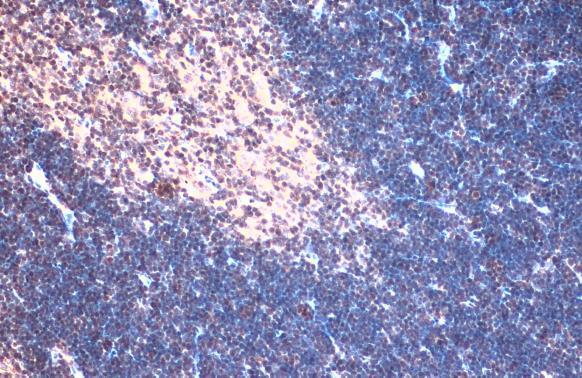
- TCF7 Polyclonal Antibody detects TCF7 protein at nucleus by immunohistochemical analysis. Sample: Paraffin-embedded mouse thymus gland. TCF7 stained by TCF7 Polyclonal Antibody (Product # PA5-78191) diluted at 1:500. Antigen Retrieval: Citrate buffer, pH 6.0, 15 min.
- Submitted by
- Invitrogen Antibodies (provider)
- Main image

- Experimental details
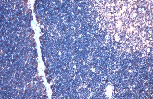
- TCF7 Polyclonal Antibody detects TCF7 protein at nucleus by immunohistochemical analysis. Sample: Paraffin-embedded rat thymus gland. TCF7 stained by TCF7 Polyclonal Antibody (Product # PA5-78191) diluted at 1:500. Antigen Retrieval: Citrate buffer, pH 6.0, 15 min.
Supportive validation
- Submitted by
- Invitrogen Antibodies (provider)
- Main image
- Experimental details
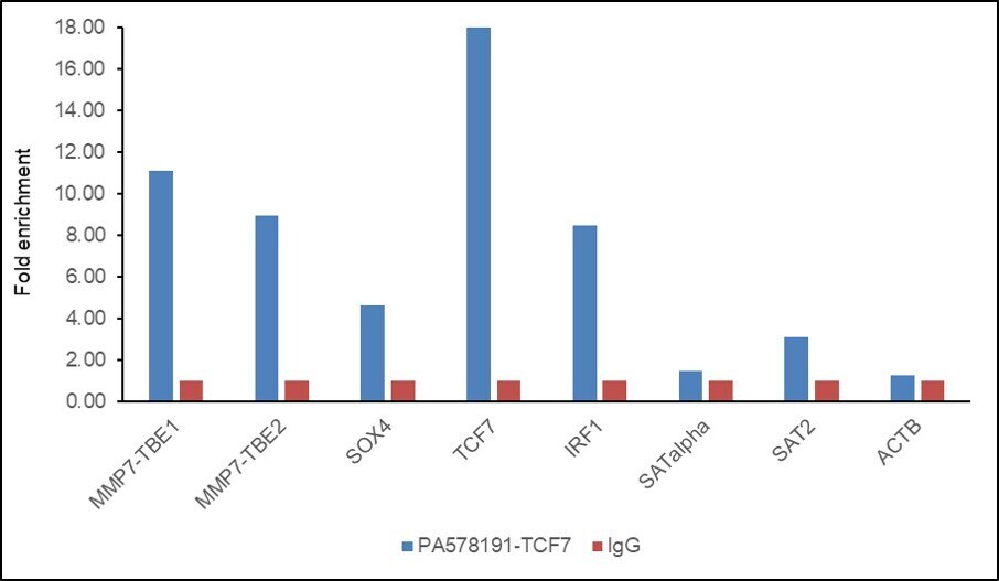
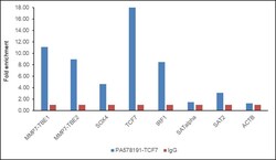
- Chromatin Immunoprecipitation (ChIP) assay of endogenous TCF7 protein using TCF7 Antibody: ChIP was performed using TCF7 Polyclonal Antibody (Product # PA5-78191, 5 µg) on sheared chromatin from HeLa cells using the MAGnify ChIP System kit (Product # 49-2024). Normal Rabbit IgG was used as a negative IP control. The purified DNA was analyzed by qPCR using primers binding to MMP7-TBE1, MMP7-TBE2, SOX4, TCF7, and IRF1 (active) and SAT alpha, SAT2 satellite repeats and Actin beta (Inactive). Data is presented as fold enrichment of the antibody signal versus the negative control IgG using the comparative CT method.