 Explore
 Validate
 Learn
Learn Western blot
 Immunoprecipitation
Antibody data
- Antibody Data
- Antigen structure
- References [0]
- Comments [0]
- Validations
- Western blot [4]
- Immunocytochemistry [1]
- Immunohistochemistry [8]
- Other assay [2]
Submit
Validation data
Reference
Comment
Report error
- Product number
- 16675-1-AP - Provider product page

- Provider
- Invitrogen Antibodies
- Product name
- AMFR Polyclonal Antibody
- Antibody type
- Polyclonal
- Antigen
- Other
- Description
- Immunogen sequence: LFHEVQRRI RRHKNYLRVV GNMEARFAVA TPEELAVNND DCAICWDSMQ AARKLPCGHL FHNSCLRSWL EQDTSCPTCR MSLNIADNNR VREEHQGENL DENLVPVAAA EGRPRLNQHN HFFHFDGSRI ASWLPSFSVE VMHTTNILGI TQASNSQLNA MAHQIQEMFP QVPYHLVLQD LQLTRSVEIT TDNILEGRIQ VPFPTQRSDS IRPALNSPVE RPSSDQEEGE TSAQTERVPL DLSPRLEETL DFGEVEVEPS EVEDFEARGS RFSKSADERQ RMLVQRKDEL LQQARKRFLN KSSEDDAASE SFLPSEGASS DPVTLRRRML AAAAERRLQK QQTS (301-643 aa encoded by B C069197)
- Reactivity
- Human, Mouse, Rat
- Host
- Rabbit
- Isotype
- IgG
- Vial size
- 150 µL
- Concentration
- 0.24 mg/mL
- Storage
- -20°C
No comments: Submit comment
Supportive validation
- Submitted by
- Invitrogen Antibodies (provider)
- Main image

- Experimental details
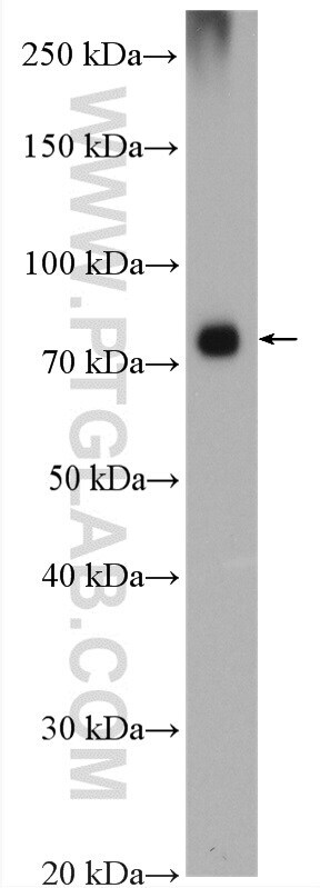
- HepG2 cells were subjected to SDS PAGE followed by western blot with 16675-1-AP (AMFR antibody) at dilution of 1:800 incubated at room temperature for 1.5 hours.

- Submitted by
- Invitrogen Antibodies (provider)
- Main image

- Experimental details
- HepG2 cells were subjected to SDS PAGE followed by western blot with 16675-1-AP (AMFR antibody) at dilution of 1:500 incubated at room temperature for 1.5 hours.

- Submitted by
- Invitrogen Antibodies (provider)
- Main image
- Experimental details
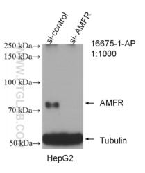
- WB result of AMFR antibody (16675-1-AP; 1:1000; incubated at room temperature for 1.5 hours) with sh-Control and sh-AMFR transfected HepG2 cells.

- Submitted by
- Invitrogen Antibodies (provider)
- Main image

- Experimental details
- MCF-7 cells were subjected to SDS PAGE followed by western blot with 16675-1-AP (AMFR antibody) at dilution of 1:600 incubated at room temperature for 1.5 hours.
Supportive validation
- Submitted by
- Invitrogen Antibodies (provider)
- Main image
- Experimental details
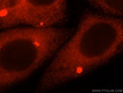
- Immunofluorescent analysis of HepG2 cells, using AMFR antibody 16675-1-AP at 1:25 dilution and Rhodamine-labeled goat anti-rabbit IGG (red).

Supportive validation
- Submitted by
- Invitrogen Antibodies (provider)
- Main image

- Experimental details
- Immunohistochemistry of paraffin-embedded human pancreas tissue slide using 16675-1-AP (AMFR antibody) at dilution of 1:200 (under 10x lens).

- Submitted by
- Invitrogen Antibodies (provider)
- Main image

- Experimental details
- Immunohistochemistry of paraffin-embedded human pancreas tissue slide using 16675-1-AP (AMFR antibody) at dilution of 1:200 (under 40x lens).

- Submitted by
- Invitrogen Antibodies (provider)
- Main image
- Experimental details
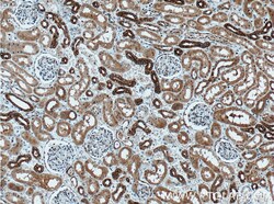
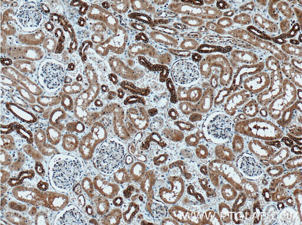
- Immunohistochemistry of paraffin-embedded human kidney tissue slide using 16675-1-AP (AMFR antibody) at dilution of 1:200 (under 10x lens) heat mediated antigen retrieved with Tris-EDTA buffer (pH 9).
- Submitted by
- Invitrogen Antibodies (provider)
- Main image

- Experimental details
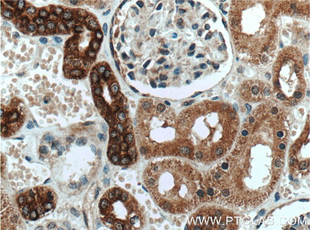
- Immunohistochemistry of paraffin-embedded human kidney tissue slide using 16675-1-AP (AMFR antibody) at dilution of 1:200 (under 40x lens) heat mediated antigen retrieved with Tris-EDTA buffer (pH 9).
- Submitted by
- Invitrogen Antibodies (provider)
- Main image
- Experimental details
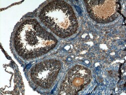
- Immunohistochemistry of paraffin-embedded mouse ovary tissue slide using 16675-1-AP (AMFR Antibody) at dilution of 1:200 (under 10x lens). heat mediated antigen retrieved with Tris-EDTA buffer (pH 9).

- Submitted by
- Invitrogen Antibodies (provider)
- Main image

- Experimental details
- Immunohistochemistry of paraffin-embedded mouse ovary tissue slide using 16675-1-AP (AMFR Antibody) at dilution of 1:200 (under 40x lens). heat mediated antigen retrieved with Tris-EDTA buffer (pH 9).
- Submitted by
- Invitrogen Antibodies (provider)
- Main image

- Experimental details
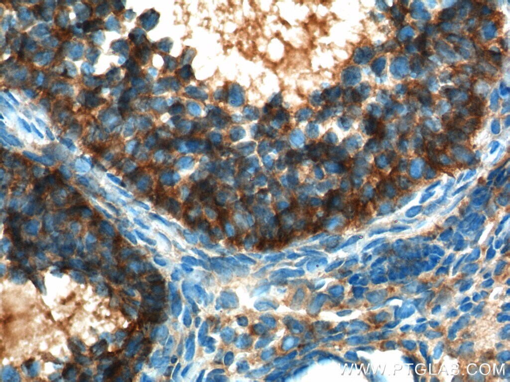
- Immunohistochemistry of paraffin-embedded human testis tissue slide using 16675-1-AP (AMFR antibody) at dilution of 1:200 (under 10x lens) heat mediated antigen retrieved with Tris-EDTA buffer (pH 9).

- Submitted by
- Invitrogen Antibodies (provider)
- Main image
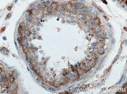
- Experimental details
- Immunohistochemistry of paraffin-embedded human testis tissue slide using 16675-1-AP (AMFR antibody) at dilution of 1:200 (under 40x lens) heat mediated antigen retrieved with Tris-EDTA buffer (pH 9).

Supportive validation
- Submitted by
- Invitrogen Antibodies (provider)
- Main image
- Experimental details
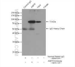
- IP result of anti-AMFR (IP:16675-1-AP, 4ug; Detection:16675-1-AP 1:300) with MCF-7 cells lysate 2400 ug.

- Submitted by
- Invitrogen Antibodies (provider)
- Main image

- Experimental details
- IP result of anti-AMFR (IP:16675-1-AP, 3 ug; Detection:16675-1-AP 1:800) with HepG2 cells lysate 2400 ug.
