 Explore
 Validate
 Learn
Learn Western blot
 Immunoprecipitation
Antibody data
- Antibody Data
- Antigen structure
- References [2]
- Comments [0]
- Validations
- Western blot [10]
- Immunohistochemistry [4]
- Flow cytometry [1]
- Other assay [2]
Submit
Validation data
Reference
Comment
Report error
- Product number
- PA5-27440 - Provider product page

- Provider
- Invitrogen Antibodies
- Product name
- KLF4 Polyclonal Antibody
- Antibody type
- Polyclonal
- Antigen
- Recombinant protein fragment
- Description
- Based on 100% sequence identity, this antibody is predicted to react with Mouse and Rat
- Reactivity
- Human, Mouse, Rat
- Host
- Rabbit
- Isotype
- IgG
- Vial size
- 100 µL
- Concentration
- 0.24 mg/mL
- Storage
- Store at 4°C short term. For long term storage, store at -20°C, avoiding freeze/thaw cycles.
Submitted references Silencing of lncRNA XIST impairs angiogenesis and exacerbates cerebral vascular injury after ischemic stroke.
KLF4 alleviates cerebral vascular injury by ameliorating vascular endothelial inflammation and regulating tight junction protein expression following ischemic stroke.
Wang C, Dong J, Sun J, Huang S, Wu F, Zhang X, Pang D, Fu Y, Li L
Molecular therapy. Nucleic acids 2021 Dec 3;26:148-160
Molecular therapy. Nucleic acids 2021 Dec 3;26:148-160
KLF4 alleviates cerebral vascular injury by ameliorating vascular endothelial inflammation and regulating tight junction protein expression following ischemic stroke.
Zhang X, Wang L, Han Z, Dong J, Pang D, Fu Y, Li L
Journal of neuroinflammation 2020 Apr 7;17(1):107
Journal of neuroinflammation 2020 Apr 7;17(1):107
No comments: Submit comment
Supportive validation
- Submitted by
- Invitrogen Antibodies (provider)
- Main image

- Experimental details
- Western blot analysis of KLF4 using 30 µg of A) A549 and B) HeLa lysate. Samples were loaded onto a 10% SDS-PAGE gel and probed with a KLF4 polyclonal antibody (Product # PA5-27440) at a dilution of 1:5000.

- Submitted by
- Invitrogen Antibodies (provider)
- Main image
- Experimental details
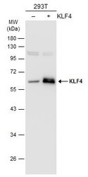
- Western Blot analysis of KLF4 was performed by separating 30 µg of non-transfected (–) and transfected (+) 293T whole cell extracts by 10% SDS-PAGE. Proteins were transferred to a membrane and probed with a KLF4 Polyclonal Antibody (Product # PA5-27440) at a dilution of 1:1000. The HRP-conjugated anti-rabbit IgG antibody was used to detect the primary antibody.

- Submitted by
- Invitrogen Antibodies (provider)
- Main image

- Experimental details
- Western Blot analysis of KLF4 was performed by separating 30 µg of HCT116 whole cell and nuclear extracts by 10% SDS-PAGE. Proteins were transferred to a membrane and probed with a KLF4 Polyclonal Antibody (Product # PA5-27440) at a dilution of 1:2000. The HRP-conjugated anti-rabbit IgG antibody was used to detect the primary antibody.

- Submitted by
- Invitrogen Antibodies (provider)
- Main image

- Experimental details
- Western Blot using KLF4 Polyclonal Antibody (Product # PA5-27440). Sample (20 µg of whole cell lysate). Lane A: mouse ESC. 12% SDS PAGE. KLF4 Polyclonal Antibody (Product # PA5-27440) diluted at 1:3,000. The HRP-conjugated anti-rabbit IgG antibody was used to detect the primary antibody.

- Submitted by
- Invitrogen Antibodies (provider)
- Main image

- Experimental details
- Western Blot analysis of KLF4 was performed by separating 30 µg of non-transfected (–) and transfected (+) 293T whole cell extracts by 10% SDS-PAGE. Proteins were transferred to a membrane and probed with a KLF4 Polyclonal Antibody (Product # PA5-27440) at a dilution of 1:1000. The HRP-conjugated anti-rabbit IgG antibody was used to detect the primary antibody.

- Submitted by
- Invitrogen Antibodies (provider)
- Main image

- Experimental details
- Western Blot using KLF4 Polyclonal Antibody (Product # PA5-27440). Sample (30 µg of whole cell lysate). Lane A: NIH-3T3. 10% SDS PAGE. KLF4 Polyclonal Antibody (Product # PA5-27440) diluted at 1:5,000. The HRP-conjugated anti-rabbit IgG antibody was used to detect the primary antibody.

- Submitted by
- Invitrogen Antibodies (provider)
- Main image

- Experimental details
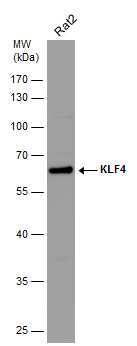
- Western Blot analysis of KLF4 was performed by separating 30 µg of various whole cell extracts by 10% SDS-PAGE. Proteins were transferred to a membrane and probed with a KLF4 Polyclonal Antibody (Product # PA5-27440) at a dilution of 1:5000 and a HRP-conjugated anti-rabbit IgG secondary antibody.

- Submitted by
- Invitrogen Antibodies (provider)
- Main image

- Experimental details
- Western Blot using KLF4 Polyclonal Antibody (Product # PA5-27440). Sample (20 µg of whole cell lysate). Lane A: human ESC. 10% SDS PAGE. KLF4 Polyclonal Antibody (Product # PA5-27440) diluted at 1:3,000. The HRP-conjugated anti-rabbit IgG antibody was used to detect the primary antibody.

- Submitted by
- Invitrogen Antibodies (provider)
- Main image

- Experimental details
- Western blot analysis of KLF4 was performed by separating 30 µg of whole cell extract by 10% SDS-PAGE. Proteins were transferred to a membrane and probed with a KLF4 Polyclonal Antibody (Product # PA5-27440) at a dilution of 1:5000. The HRP-conjugated anti-rabbit IgG antibody was used to detect the primary antibody.
- Submitted by
- Invitrogen Antibodies (provider)
- Main image

- Experimental details
- Western Blot using KLF4 Polyclonal Antibody (Product # PA5-27440). Various whole cell extracts (30 µg) were separated by 10% SDS-PAGE, and the membrane was blotted with KLF4 Polyclonal Antibody (Product # PA5-27440) diluted at 1:5,000. The HRP-conjugated anti-rabbit IgG antibody was used to detect the primary antibody.

Supportive validation
- Submitted by
- Invitrogen Antibodies (provider)
- Main image

- Experimental details
- KLF4 Polyclonal Antibody detects KLF4 protein at cytoplasm and nucleus by immunohistochemical analysis. Sample: Paraffin-embedded mouse lung. KLF4 stained by KLF4 Polyclonal Antibody (Product # PA5-27440) diluted at 1:1,000. Antigen Retrieval: Citrate buffer, pH 6.0, 15 min.

- Submitted by
- Invitrogen Antibodies (provider)
- Main image

- Experimental details
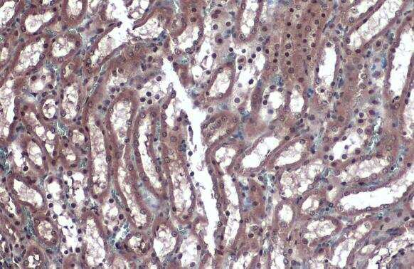
- KLF4 Polyclonal Antibody detects KLF4 protein at cytoplasm and nucleus by immunohistochemical analysis. Sample: Paraffin-embedded rat kidney. KLF4 stained by KLF4 Polyclonal Antibody (Product # PA5-27440) diluted at 1:1,000. Antigen Retrieval: Citrate buffer, pH 6.0, 15 min.
- Submitted by
- Invitrogen Antibodies (provider)
- Main image

- Experimental details
- KLF4 Polyclonal Antibody detects KLF4 protein at cytoplasm and nucleus by immunohistochemical analysis. Sample: Paraffin-embedded rat lung. KLF4 stained by KLF4 Polyclonal Antibody (Product # PA5-27440) diluted at 1:1,000. Antigen Retrieval: Citrate buffer, pH 6.0, 15 min.

Supportive validation
- Submitted by
- Invitrogen Antibodies (provider)
- Main image

- Experimental details
- Flow cytometry on human embryonic stem cells, staining with KLF4 Polyclonal Antibody (Product # PA5-27440) at 1:100 dilution (purple) or rabbit IgG (black).

Supportive validation
- Submitted by
- Invitrogen Antibodies (provider)
- Main image

- Experimental details
- Figure 5 Overexpression of Itgalpha5 or KLF4 reverses the effect of lncRNA XIST on the migration and tube formation and the expression of three CAMs, p-NF-kappaB, and TJPs of bEnd3 cells under OGD/R conditions (A) Representative images of western blot for the expression of Itgalpha5 in bEnd3 cells co-transfected with si-XIST and Itgalpha5 overexpression plasmid at 24 h restoration from OGD. (B) Bar graphs show the quantitative analyses of western blots as ratios of Itgalpha5/beta-actin (n = 4 per experimental group). (C) Representative images of cell migration and capillary tube formation of bEnd3 cells co-transfected with si-XIST and Itgalpha5 overexpression plasmid after OGD/R treatment. Scale bar, 400 mum for migration; 500 mum for tube formation. (D and E) Quantification of cell migration (D) and tube formation (E) of bEnd3 cells (n = 4 per experimental group). (F) Representative images of western blot for the expression of KLF4, three CAMs (E-selectin, VCAM-1, and ICAM-1), p-NF-kappaB, and TJPs (Claudin-5 and ZO-1) in bEnd3 cells co-transfected with si-XIST and KLF4 overexpression plasmid at 24 h restoration from OGD. (G-M) Bar graphs show the quantitative analyses of western blots as ratios of KLF4/beta-actin (G), E-selectin/beta-actin (H), VCAM-1/beta-actin (I), ICAM-1/beta-actin (J), p-NF-kappaB/total NF-kappaB (K), Claudin-5/beta-actin (L), and ZO-1/beta-actin (M) (n = 4 per experimental group). Note that bEnd3 cells co-transfected with si-XIST and Itgalpha5 displaye

- Submitted by
- Invitrogen Antibodies (provider)
- Main image

- Experimental details
- Figure 6 lncRNA XIST alleviates a vascular endothelial inflammation response and regulates migration and tube formation of bEnd3 cells under OGD/R conditions by targeting miR-92a (A) Representative images of western blot for the expression of Itgalpha5, KLF4, E-selectin, VCAM-1, ICAM-1, p-NF-kappaB, Claudin-5, and ZO-1 in bEnd3 cells co-transfected with the si-Ctl or si-XIST and/or miR-92a inhibitor (miR-92a-I) at 24 h restoration from OGD. (B-I) Bar graphs show the quantitative analyses of western blots as ratios of Itgalpha5/beta-actin (B), KLF4/beta-actin (C), E-selectin/beta-actin (D), VCAM-1/beta-actin (E), ICAM-1/beta-actin (F), p-NF-kappaB/total NF-kappaB (G), Claudin-5/beta-actin (H), and ZO-1/beta-actin (I) (n = 4 per experimental group). Note that the expressions of Itgalpha5, KLF4, and TJPs (Claudin-5 and ZO-1) in the si-XIST + NC inhibitor (NC-I)-treated bEnd3 cells markedly decreased, but the expressions of three CAMs including E-selectin, VCAM-1, and ICAM-1 and the p-NF-kappaB were significantly elevated relative to the si-Ctl + NC-I-treated group at 24 h restoration from OGD. However, these effects were significantly rescued by inhibiting the levels of miR-92a in the bEnd3 cells. *p < 0.05, **p < 0.01, and ***p < 0.001. (J) Representative images of cell migration and capillary tube formation of bEnd3 cells transfected with si-Ctl or si-XIST and/or miR-92a-I after OGD/R treatment. Scale bar, 400 mum for migration; 500 mum for tube formation. (K and L) Quantifica
