 Explore
 Validate
 Learn
Learn Western blot
 Immunocytochemistry
Antibody data
- Antibody Data
- Antigen structure
- References [0]
- Comments [0]
- Validations
- Immunocytochemistry [9]
- Immunohistochemistry [5]
- Flow cytometry [2]
Submit
Validation data
Reference
Comment
Report error
- Product number
- MA5-34642 - Provider product page

- Provider
- Invitrogen Antibodies
- Product name
- MATR3 Recombinant Rabbit Monoclonal Antibody (JU93-43)
- Antibody type
- Monoclonal
- Antigen
- Synthetic peptide
- Description
- Positive Control: Daudi, mouse brain tissue lysate, Hela, LOVO, SH-SY-5Y, rat brain tissue, human tonsil tissue, human lung cancer tissue, human liver tissue, mouse testis tissue.
- Reactivity
- Human
- Host
- Rabbit
- Isotype
- IgG
- Antibody clone number
- JU93-43
- Vial size
- 100 μL
- Concentration
- 1 mg/mL
- Storage
- -20°C, Avoid Freeze/Thaw Cycles, store in dark
No comments: Submit comment
Supportive validation
- Submitted by
- Invitrogen Antibodies (provider)
- Main image

- Experimental details
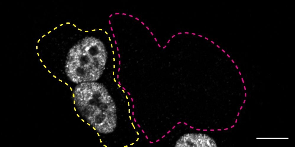
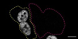
- Immunofluorescence of MATR3 was performed using HAP1 wild-type and MATR3 KO cells that were transfected with a green or a far-red fluorescent dye, respectively. Post-transfection, WT and KO cells were mixed and plated to a 1:1 ratio on coverslips as a mosaic and incubated for 24 hrs. Cells were fixed in 4% PFA (in PBS) for 15 min; cells were permeabilized with 0.1% Triton X-100 for 10 min at RT and blocked with PBS with 5% BSA, 5% goat serum, and 0.01% Triton X-100 for 30 min. Cells were stained with the MATR3 recombinant monoclonal antibody (Product # MA5-34642) at a 1:1,000 dilution overnight at 4°C. Secondary antibody incubation was performed using 1 µg/mL of Goat anti-Rabbit IgG (H+L) Highly Cross-Adsorbed Secondary Antibody, Alexa Fluor 555 antibody (Product # A21429) for 1 hr. Imaging was performed with a 40X oil objective and analysis was performed using Image J. Cell image represents a single focal plane; WT and KO cells are outlined with a yellow (WT) or magenta (KO) dashed line. Data courtesy of YCharOS Inc., an open science company with the mission of characterizing commercially available antibodies using knockout validation.
- Submitted by
- Invitrogen Antibodies (provider)
- Main image

- Experimental details
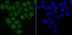
- Immunofluorescent analysis of Matrin 3 in SH-SY-5Y cells (green). Samples were fixed in paraformaldehyde and permeabilised with 0.25% Triton X100/PBS, incubated with Matrin 3 monoclonal antibody (Product # MA5-34642), followed by DAPI (blue).

- Submitted by
- Invitrogen Antibodies (provider)
- Main image
- Experimental details
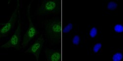
- Immunofluorescent analysis of Matrin 3 in LOVO cells (green). Samples were fixed in paraformaldehyde and permeabilised with 0.25% Triton X100/PBS, incubated with Matrin 3 monoclonal antibody (Product # MA5-34642), followed by DAPI (blue).

- Submitted by
- Invitrogen Antibodies (provider)
- Main image

- Experimental details
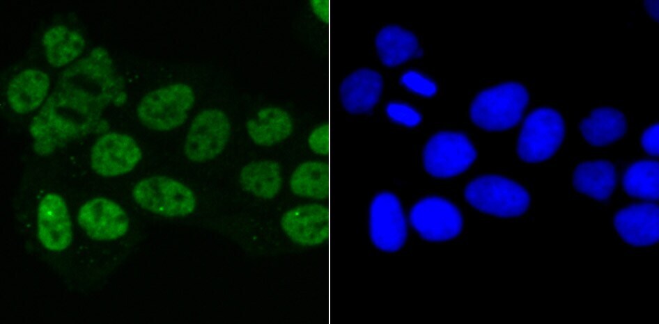
- Immunofluorescent analysis of Matrin 3 in HeLa cells (green). Samples were fixed in paraformaldehyde and permeabilised with 0.25% Triton X100/PBS, incubated with Matrin 3 monoclonal antibody (Product # MA5-34642), followed by DAPI (blue).
- Submitted by
- Invitrogen Antibodies (provider)
- Main image

- Experimental details
- Immunofluorescent analysis of Matrin 3 in HeLa cells (green). Samples were fixed in paraformaldehyde and permeabilised with 0.25% Triton X100/PBS, incubated with Matrin 3 monoclonal antibody (Product # MA5-34642), followed by DAPI (blue).

- Submitted by
- Invitrogen Antibodies (provider)
- Main image

- Experimental details
- Immunofluorescent analysis of Matrin 3 in LOVO cells (green). Samples were fixed in paraformaldehyde and permeabilised with 0.25% Triton X100/PBS, incubated with Matrin 3 monoclonal antibody (Product # MA5-34642), followed by DAPI (blue).

- Submitted by
- Invitrogen Antibodies (provider)
- Main image
- Experimental details
- Immunofluorescent analysis of Matrin 3 in SH-SY-5Y cells (green). Samples were fixed in paraformaldehyde and permeabilised with 0.25% Triton X100/PBS, incubated with Matrin 3 monoclonal antibody (Product # MA5-34642), followed by DAPI (blue).

- Submitted by
- Invitrogen Antibodies (provider)
- Main image
- Experimental details
- Immunofluorescence of MATR3 was performed using HAP1 wild-type and MATR3 KO cells that were transfected with a green or a far-red fluorescent dye, respectively. Post-transfection, WT and KO cells were mixed and plated to a 1:1 ratio on coverslips as a mosaic and incubated for 24 hrs. Cells were fixed in 4% PFA (in PBS) for 15 min; cells were permeabilized with 0.1% Triton X-100 for 10 min at RT and blocked with PBS with 5% BSA, 5% goat serum, and 0.01% Triton X-100 for 30 min. Cells were stained with the MATR3 recombinant monoclonal antibody (Product # MA5-34642) at a 1:1,000 dilution overnight at 4°C. Secondary antibody incubation was performed using 1 µg/mL of Goat anti-Rabbit IgG (H+L) Highly Cross-Adsorbed Secondary Antibody, Alexa Fluor 555 antibody (Product # A21429) for 1 hr. Imaging was performed with a 40X oil objective and analysis was performed using Image J. Cell image represents a single focal plane; WT and KO cells are outlined with a yellow (WT) or magenta (KO) dashed line. Data courtesy of YCharOS Inc., an open science company with the mission of characterizing commercially available antibodies using knockout validation.

- Submitted by
- Invitrogen Antibodies (provider)
- Main image
- Experimental details
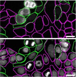
- Immunofluorescence of matrin-3 was performed using HAP1 WT and MATR3 KO cells that were labeled with a green or a far-red fluorescent dye, respectively. Post-labeling, WT and KO cells were mixed and plated to a 1:1 ratio in a 96-well plate with an optically clear flat bottom as a mosaic and incubated for 24 hrs. Cells were fixed in 4% PFA (in PBS) for 15 min at RT; cells were permeabilized with 0.1% Triton X-100 for 10 min at RT and blocked with PBS containing 5% BSA, 5% goat serum, and 0.01% Triton X-100 for 30 min at RT. Cells were stained with MATR3 Recombinant Rabbit Monoclonal Antibody (JU93-43) (Product # MA5-34642) at a 1:1,000 dilution overnight at 4 degrees Celcius. Secondary antibody incubation was performed using 1 µg/mL of Goat anti-Rabbit IgG (H+L) Highly Cross-Adsorbed Secondary Antibody, Alexa Fluor™ 555 (Product # A-21429) together with DAPI for 1 hr at RT. Imaging was performed with a 20X water immersion objective. Representative images where WT and KO cells are outlined with a green (WT) or magenta (KO) line, respectively, are shown. The top and bottom panels show antibody and DAPI stainings, respectively. Scale bar = 10 μm. Data courtesy of YCharOS Inc., an open science company with the mission of characterizing commercially available antibodies using knockout validation.

Supportive validation
- Submitted by
- Invitrogen Antibodies (provider)
- Main image
- Experimental details
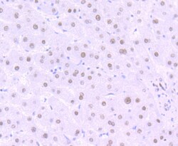
- Immunohistochemistry analysis of Matrin 3 in paraffin-embedded human liver tissue. Samples were incubated with Matrin 3 monoclonal antibody (Product # MA5-34642), and followed by hematoxylin.

- Submitted by
- Invitrogen Antibodies (provider)
- Main image
- Experimental details
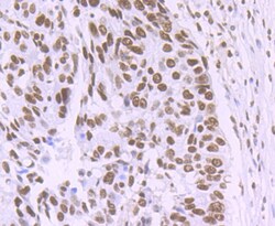
- Immunohistochemistry analysis of Matrin 3 in paraffin-embedded human lung cancer tissue. Samples were incubated with Matrin 3 monoclonal antibody (Product # MA5-34642), and followed by hematoxylin.

- Submitted by
- Invitrogen Antibodies (provider)
- Main image
- Experimental details
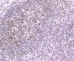
- Immunohistochemistry analysis of Matrin 3 in paraffin-embedded human tonsil tissue. Samples were incubated with Matrin 3 monoclonal antibody (Product # MA5-34642), and followed by hematoxylin.

- Submitted by
- Invitrogen Antibodies (provider)
- Main image
- Experimental details
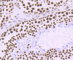
- Immunohistochemistry analysis of Matrin 3 in paraffin-embedded mouse testis tissue. Samples were incubated with Matrin 3 monoclonal antibody (Product # MA5-34642), and followed by hematoxylin.

- Submitted by
- Invitrogen Antibodies (provider)
- Main image
- Experimental details
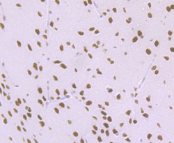
- Immunohistochemistry analysis of Matrin 3 in paraffin-embedded rat brain tissue. Samples were incubated with Matrin 3 monoclonal antibody (Product # MA5-34642), and followed by hematoxylin.

Supportive validation
- Submitted by
- Invitrogen Antibodies (provider)
- Main image

- Experimental details
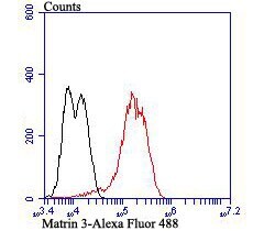
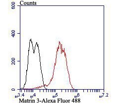
- Flow cytometry of Matrin 3 in LOVO cells (red) compared with an unlabelled control (cells without incubation with primary antibody; black). Samples were incubated with Matrin 3 monoclonal antibody (Product # MA5-34642) at a dilution of 1:100, followed by Alexa Fluor 488-conjugated goat anti-rabbit IgG.
- Submitted by
- Invitrogen Antibodies (provider)
- Main image
- Experimental details
- Flow cytometry of Matrin 3 in LOVO cells (red) compared with an unlabelled control (cells without incubation with primary antibody; black). Samples were incubated with Matrin 3 monoclonal antibody (Product # MA5-34642) at a dilution of 1:100, followed by Alexa Fluor 488-conjugated goat anti-rabbit IgG.
