 Explore
 Validate
 Learn
Learn Western blot
 Immunocytochemistry
Antibody data
- Antibody Data
- Antigen structure
- References [1]
- Comments [0]
- Validations
- Western blot [5]
- Immunoprecipitation [1]
- Immunohistochemistry [8]
Submit
Validation data
Reference
Comment
Report error
- Product number
- NBP2-16309 - Provider product page

- Provider
- Novus Biologicals
- Product name
- Rabbit Polyclonal eIF4GII Antibody
- Antibody type
- Polyclonal
- Description
- Immunogen affinity purified.
- Reactivity
- Human, Mouse, Rat
- Host
- Rabbit
- Isotype
- IgG
- Vial size
- 0.1 ml
- Storage
- Aliquot and store at -20C or -80C. Avoid freeze-thaw cycles.
Submitted references A Translation-Activating Function of MIWI/piRNA during Mouse Spermiogenesis.
Dai P, Wang X, Gou LT, Li ZT, Wen Z, Chen ZG, Hua MM, Zhong A, Wang L, Su H, Wan H, Qian K, Liao L, Li J, Tian B, Li D, Fu XD, Shi HJ, Zhou Y, Liu MF
Cell 2019 Dec 12;179(7):1566-1581.e16
Cell 2019 Dec 12;179(7):1566-1581.e16
No comments: Submit comment
Supportive validation
- Submitted by
- Novus Biologicals (provider)
- Main image

- Experimental details
- Western Blot: eIF4GII Antibody [NBP2-16309] - Sample (30 ug of whole cell lysate) A: JC B: BCL-1 5% SDS PAGE gel, diluted at 1:5000.

- Submitted by
- Novus Biologicals (provider)
- Main image
- Experimental details
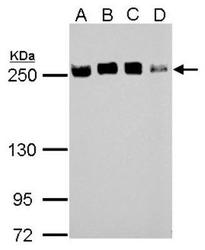
- Western Blot: eIF4GII Antibody [NBP2-16309] - Sample (30 ug of whole cell lysate) A: 293T B: A431 C: HeLa D: HepG2 5% SDS PAGE gel, diluted at 1:10000.

- Submitted by
- Novus Biologicals (provider)
- Main image
- Experimental details
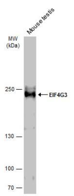
- Western Blot: eIF4GII Antibody [NBP2-16309] - A: Marker. B: mouse testes. 6% SDS-PAGE gel, antibody diluted 1:10000. This image was submitted via customer review.

- Submitted by
- Novus Biologicals (provider)
- Main image

- Experimental details
- Western Blot: eIF4GII Antibody [NBP2-16309] - Mouse tissue extract (50 ug) was separated by 5% SDS-PAGE, and the membrane was blotted with eIF4GII antibody diluted at 1:1000. HRP-conjugated anti-rabbit IgG antibody was used to detect the primary antibody.
- Submitted by
- Novus Biologicals (provider)
- Main image

- Experimental details
- Western Blot: eIF4GII Antibody [NBP2-16309] - Various whole cell extracts (30 ug) were separated by 5% SDS-PAGE, and the membrane was blotted with eIF4GII antibody diluted at 1:10000.

Supportive validation
- Submitted by
- Novus Biologicals (provider)
- Main image
- Experimental details
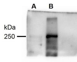
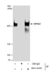
- Immunoprecipitation: eIF4GII Antibody [NBP2-16309] - Immunoprecipitation of EIF4G3 protein from 293T whole cell extracts using 5 ug of EIF4G3 antibody [N1], N-term. Western blot analysis was performed using EIF4G3 ntibody [N1], N-term. EasyBlot anti-Rabbit IgG was used as a secondary reagent

Supportive validation
- Submitted by
- Novus Biologicals (provider)
- Main image

- Experimental details
- Immunohistochemistry-Paraffin: eIF4GII Antibody [NBP2-16309] - Immunohistochemical analysis of paraffin-embedded AGS xenograft, using antibody at 1:500 dilution.
- Submitted by
- Novus Biologicals (provider)
- Main image
- Experimental details
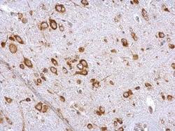
- Immunohistochemistry-Paraffin: eIF4GII Antibody [NBP2-16309] - Paraffin-embedded mouse brain stem. EIF4G3 antibody [N1], N-term dilution: 1:500.
- Submitted by
- Novus Biologicals (provider)
- Main image
- Experimental details
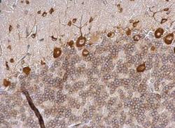
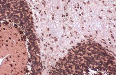
- Immunohistochemistry-Paraffin: eIF4GII Antibody [NBP2-16309] - Paraffin-embedded rat hind brain. EIF4G3 antibody [N1], N-term dilution: 1:500.

- Submitted by
- Novus Biologicals (provider)
- Main image

- Experimental details
- Immunohistochemistry-Paraffin: eIF4GII Antibody [NBP2-16309] - Paraffin-embedded mouse cervix. EIF4G3 antibody [N1], N-term dilution: 1:500.

- Submitted by
- Novus Biologicals (provider)
- Main image

- Experimental details
- Immunohistochemistry-Frozen: eIF4GII Antibody [NBP2-16309] - eIF4GII antibody detects eIF4GII protein expression by immunohistochemical analysis. Sample: Frozen-sectioned adult mouse cerebellum. Green: eIF4GII protein stained by eIF4GII antibody diluted at 1:250. Red: NeuN, stained by NeuN antibody diluted at 1:500. Blue: Fluoroshield with DAPI.

- Submitted by
- Novus Biologicals (provider)
- Main image
- Experimental details
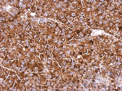
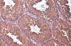
- Immunohistochemistry-Paraffin: eIF4GII Antibody [NBP2-16309] - eIF4GII antibody [N1], N-term detects eIF4GII protein at cytoplasm by immunohistochemical analysis. Sample: Paraffin-embedded mouse testis. eIF4GII stained by eIF4GII diluted at 1:500. Antigen Retrieval: Citrate buffer, pH 6.0, 15 min.

- Submitted by
- Novus Biologicals (provider)
- Main image

- Experimental details
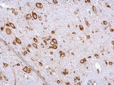
- Immunohistochemistry-Paraffin: eIF4GII Antibody [NBP2-16309] - eIF4GII antibody detects eIF4GII protein at cytoplasm by immunohistochemical analysis. Sample: Paraffin-embedded mouse brain. eIF4GII stained by eIF4GII antibody diluted at 1:500. Antigen Retrieval: Citrate buffer, pH 6.0, 15 min.
- Submitted by
- Novus Biologicals (provider)
- Main image

- Experimental details
- Immunohistochemistry-Paraffin: eIF4GII Antibody [NBP2-16309] - eIF4GII antibody detects eIF4GII protein at cytoplasm by immunohistochemical analysis. Sample: Paraffin-embedded rat brain. eIF4GII stained by eIF4GII antibody diluted at 1:500. Antigen Retrieval: Citrate buffer, pH 6.0, 15 min.
