Explore
 Validate
 Learn
Learn Western blot
Antibody data
- Antibody Data
- Antigen structure
- References [4]
- Comments [0]
- Validations
- Western blot [2]
- ELISA [1]
- Immunocytochemistry [1]
- Immunoprecipitation [1]
- Immunohistochemistry [1]
Submit
Validation data
Reference
Comment
Report error
- Product number
- H00010963-M01 - Provider product page

- Provider
- Novus Biologicals
- Proper citation
- Novus Cat#H00010963-M01, RRID:AB_548439
- Product name
- Mouse Monoclonal STI1 Antibody
- Antibody type
- Monoclonal
- Description
- IgG purified. STIP1 - stress-induced-phosphoprotein 1 (Hsp70/Hsp90-organizing protein)
- Reactivity
- Human, Mouse
- Host
- Mouse
- Isotype
- IgG
- Vial size
- 0.1 mg
- Storage
- Aliquot and store at -20C or -80C. Avoid freeze-thaw cycles.
Submitted references Distinct roles of molecular chaperones HSP90α and HSP90β in the biogenesis of KCNQ4 channels.
Stress-induced phosphoprotein 1 as a secreted biomarker for human ovarian cancer promotes cancer cell proliferation.
Proteomic analysis of protein complexes in human SH-SY5Y neuroblastoma cells by using blue-native gel electrophoresis: an increase in lamin A/C associated with heat shock protein 90 in response to 6-hydroxydopamine-induced oxidative stress.
Novel breast cancer metastasis-associated proteins.
Gao Y, Yechikov S, Vazquez AE, Chen D, Nie L
PloS one 2013;8(2):e57282
PloS one 2013;8(2):e57282
Stress-induced phosphoprotein 1 as a secreted biomarker for human ovarian cancer promotes cancer cell proliferation.
Wang TH, Chao A, Tsai CL, Chang CL, Chen SH, Lee YS, Chen JK, Lin YJ, Chang PY, Wang CJ, Chao AS, Chang SD, Chang TC, Lai CH, Wang HS
Molecular & cellular proteomics : MCP 2010 Sep;9(9):1873-84
Molecular & cellular proteomics : MCP 2010 Sep;9(9):1873-84
Proteomic analysis of protein complexes in human SH-SY5Y neuroblastoma cells by using blue-native gel electrophoresis: an increase in lamin A/C associated with heat shock protein 90 in response to 6-hydroxydopamine-induced oxidative stress.
Nakamura M, Morisawa H, Imajoh-Ohmi S, Takamura C, Fukuda H, Toda T
Experimental gerontology 2009 Jun-Jul;44(6-7):375-82
Experimental gerontology 2009 Jun-Jul;44(6-7):375-82
Novel breast cancer metastasis-associated proteins.
Ho J, Kong JW, Choong LY, Loh MC, Toy W, Chong PK, Wong CH, Wong CY, Shah N, Lim YP
Journal of proteome research 2009 Feb;8(2):583-94
Journal of proteome research 2009 Feb;8(2):583-94
No comments: Submit comment
Supportive validation
- Submitted by
- Novus Biologicals (provider)
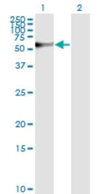
- Main image

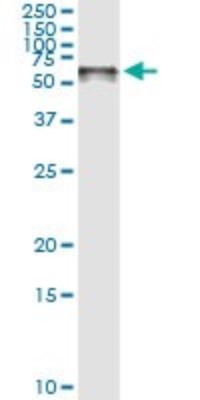
- Experimental details
- Western Blot: STI1 Antibody (4B6) [H00010963-M01] - Analysis of STIP1 expression in transfected 293T cell line by STIP1 monoclonal antibody (M01), clone 4B6.Lane 1: STIP1 transfected lysate(62.6 KDa).Lane 2: Non-transfected lysate.
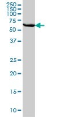
- Submitted by
- Novus Biologicals (provider)
- Main image

- Experimental details
- Western Blot: STI1 Antibody (4B6) [H00010963-M01] - STIP1 monoclonal antibody (M01), clone 4B6 Analysis of STIP1 expression in HeLa.
Supportive validation
- Submitted by
- Novus Biologicals (provider)
- Main image

- Experimental details
- Sandwich ELISA: STI1 Antibody (4B6) [H00010963-M01] - Detection limit for recombinant GST tagged STIP1 is approximately 0.1ng/ml as a capture antibody.
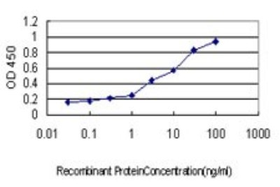
Supportive validation
- Submitted by
- Novus Biologicals (provider)
- Main image
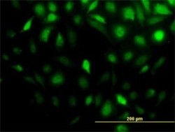
- Experimental details
- Immunocytochemistry/Immunofluorescence: STI1 Antibody (4B6) [H00010963-M01] - Analysis of monoclonal antibody to STIP1 on HeLa cell. Antibody concentration 10 ug/ml.

Supportive validation
- Submitted by
- Novus Biologicals (provider)
- Main image

- Experimental details
- Immunoprecipitation: STI1 Antibody (4B6) [H00010963-M01] - Analysis of STIP1 transfected lysate using anti-STIP1 monoclonal antibody and Protein A Magnetic Bead, and immunoblotted with STIP1 MaxPab rabbit polyclonal antibody.
Supportive validation
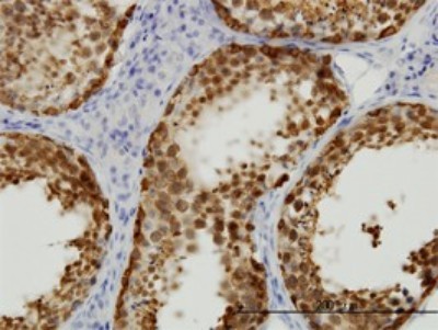
- Submitted by
- Novus Biologicals (provider)
- Main image

- Experimental details
- Immunohistochemistry-Paraffin: STI1 Antibody (4B6) [H00010963-M01] - Analysis of monoclonal antibody to STIP1 on formalin-fixed paraffin-embedded human testis. Antibody concentration 0.3 ug/ml.