Explore
 Validate
 Learn
Learn Western blot
Antibody data
- Antibody Data
- Antigen structure
- References [3]
- Comments [0]
- Validations
- Western blot [2]
- ELISA [1]
- Immunocytochemistry [1]
- Immunohistochemistry [1]
- Flow cytometry [1]
Submit
Validation data
Reference
Comment
Report error
- Product number
- H00010963-M35 - Provider product page

- Provider
- Novus Biologicals
- Proper citation
- Novus Cat#H00010963-M35, RRID:AB_2198176
- Product name
- Mouse Monoclonal STI1 Antibody
- Antibody type
- Monoclonal
- Description
- IgG purified.
- Reactivity
- Human
- Host
- Mouse
- Isotype
- IgG
- Vial size
- 0.1 mg
- Storage
- Aliquot and store at -20C or -80C. Avoid freeze-thaw cycles.
Submitted references Tumor stress-induced phosphoprotein1 (STIP1) as a prognostic biomarker in ovarian cancer.
Immunohistological analysis of stress-induced phosphoprotein 1 in ovarian cancer patients with low serum cancer antigen 125 levels.
Stress-induced phosphoprotein 1 as a secreted biomarker for human ovarian cancer promotes cancer cell proliferation.
Chao A, Lai CH, Tsai CL, Hsueh S, Hsueh C, Lin CY, Chou HH, Lin YJ, Chen HW, Chang TC, Wang TH
PloS one 2013;8(2):e57084
PloS one 2013;8(2):e57084
Immunohistological analysis of stress-induced phosphoprotein 1 in ovarian cancer patients with low serum cancer antigen 125 levels.
Chao A, Lee LY, Hsueh C, Lin CY, Tsai CL, Chao AS, Lin CT, Chou HH, Chang TC, Wang TH
Taiwanese journal of obstetrics & gynecology 2013 Jun;52(2):185-91
Taiwanese journal of obstetrics & gynecology 2013 Jun;52(2):185-91
Stress-induced phosphoprotein 1 as a secreted biomarker for human ovarian cancer promotes cancer cell proliferation.
Wang TH, Chao A, Tsai CL, Chang CL, Chen SH, Lee YS, Chen JK, Lin YJ, Chang PY, Wang CJ, Chao AS, Chang SD, Chang TC, Lai CH, Wang HS
Molecular & cellular proteomics : MCP 2010 Sep;9(9):1873-84
Molecular & cellular proteomics : MCP 2010 Sep;9(9):1873-84
No comments: Submit comment
Supportive validation
- Submitted by
- Novus Biologicals (provider)
- Main image

- Experimental details
- Western Blot: STI1 Antibody (2E11) [H00010963-M35] - Analysis of STIP1 expression in transfected 293T cell line by STIP1 monoclonal antibody (M35), clone 2E11. Lane 1: STIP1 transfected lysate (Predicted MW: 62.6 KDa). Lane 2: Non-transfected lysate.

- Submitted by
- Novus Biologicals (provider)
- Main image

- Experimental details
- Western Blot: STI1 Antibody (2E11) [H00010963-M35] - Analysis of STIP1 expression in MCF-7.

Supportive validation
- Submitted by
- Novus Biologicals (provider)
- Main image

- Experimental details
- Sandwich ELISA: STI1 Antibody (2E11) [H00010963-M35] - Detection limit for recombinant GST tagged STIP1 is 0.1 ng/ml as a capture antibody.

Supportive validation
- Submitted by
- Novus Biologicals (provider)
- Main image

- Experimental details
- Immunocytochemistry/Immunofluorescence: STI1 Antibody (2E11) [H00010963-M35] - Analysis of monoclonal antibody to STIP1 on HeLa cell. Antibody concentration 10 ug/ml

Supportive validation
- Submitted by
- Novus Biologicals (provider)
- Main image

- Experimental details
- Immunohistochemistry-Paraffin: STI1 Antibody (2E11) [H00010963-M35] - Analysis of monoclonal antibody to STIP1 on formalin-fixed paraffin-embedded human ovarian cancer. Antibody concentration 0.75 ug/ml

Supportive validation
- Submitted by
- Novus Biologicals (provider)
- Main image

- Experimental details
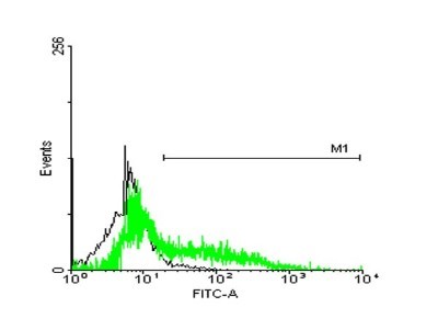
- Flow Cytometry: STI1 Antibody (2E11) [H00010963-M35] - Analysis of HeLa cells stained with STIP1 monoclonal antibody clone 2E11 (Green) and non-stained HeLa cells (Black) as negative control.