 Explore
 Validate
 Learn
Learn Immunohistochemistry
Antibody data
- Antibody Data
- Antigen structure
- References [1]
- Comments [0]
- Validations
- Immunohistochemistry [1]
- Other assay [6]
Submit
Validation data
Reference
Comment
Report error
- Product number
- PA5-41479 - Provider product page

- Provider
- Invitrogen Antibodies
- Product name
- HOXC6 Polyclonal Antibody
- Antibody type
- Polyclonal
- Antigen
- Synthetic peptide
- Reactivity
- Human, Mouse
- Host
- Rabbit
- Isotype
- IgG
- Vial size
- 100 µL
- Concentration
- 0.5 mg/mL
- Storage
- -20° C, Avoid Freeze/Thaw Cycles
Submitted references HomeoboxC6 affects the apoptosis of human vascular endothelial cells and is involved in atherosclerosis.
Long X, You G, Wu Q, Zhou Y, Xiao Y, Yu F, Deng S, Mo R, Song F, Huang J, Tian M
Journal of cellular physiology 2021 Mar;236(3):1913-1925
Journal of cellular physiology 2021 Mar;236(3):1913-1925
No comments: Submit comment
Supportive validation
- Submitted by
- Invitrogen Antibodies (provider)
- Main image

- Experimental details
- Immunofluorescence analysis of mouse spinal cord cells using an anti-HOXC6 polyclonal antibody (Product # PA5-41479).

Supportive validation
- Submitted by
- Invitrogen Antibodies (provider)
- Main image
- Experimental details
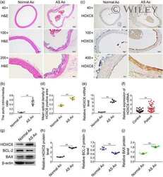
- 1 Figure Abnormal expression of HOXC6, BCL-2, and BAX in aortic wall of AS rats or PBMs of patients with CHD. (a,b) H&E staining showing the structural characteristics of normal and atherosclerotic aorta of rats and analysis of the intima/media ratio. (c,d) Immunohistochemical staining showing the expression of HOXC6 protein in normal and atherosclerotic aorta of rats and analysis of the mean optical density of HOXC6-positive area. (e) Relative HOXC6 mRNA expression in normal and atherosclerotic aorta of rats was determined by RT-qPCR. (f) Relative HOXC6 mRNA expression in PBMs of control population and patients with CHD was detected by RT-qPCR. (g-j) Western blot analysis showing the expressions of HOXC6, BCL-2, and BAX and densitometric analysis of each protein/beta-actin. AS, atherosclerosis; Bax, Bcl-2 associated X; Bcl-2, B-cell lymphoma 2; CHD, coronary heart disease; HOXC6, homeboxC6; mRNA, messenger RNA; PBM, peripheral blood monocyte; RT-qPCR, quantitative reverse transcription polymerase chain reaction. ** p < .01 versus normal group

- Submitted by
- Invitrogen Antibodies (provider)
- Main image

- Experimental details
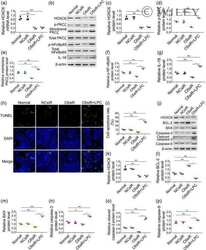
- 2 Figure HOXC6 -siRNA inhibits the apoptosis of HVECs cultured normally or treated with ox-LDL. Cells were treated with 50 mug/mL ox-LDL for 12 hr followed by transfection with siRNAs for 24-48 hr. (a,b) TUNEL assay showing the apoptosis of HVECs and analysis of the cell apoptosis rate. (c) Relative HOXC6 mRNA expression was identified by RT-qPCR. (d-j) Western blot analysis showing the expressions of HOXC6 and apoptosis-related proteins and densitometric analysis of each protein/beta-actin. HOXC6, homeboxC6; HVEC, human vascular endothelial cell; ox-LDL, oxidized low-density lipoprotein; siRNA, small interfering RNA; TUNEL, terminal deoxynucleotidyl transferase-mediated dUTP nick end labeling. * p < .05, ** p < .01 versus normal group; p < .01 versus OX-L group

- Submitted by
- Invitrogen Antibodies (provider)
- Main image

- Experimental details
- 3 Figure HOXC6 -overexpressed plasmid promotes the apoptosis of HVECs. Cells were transfected with plasmid vectors for 48-72 hr. (a,b) TUNEL assay showing the apoptosis of HVECs and analysis of the cell apoptosis rate. (c) Relative expression of HOXC6 mRNA in different groups was determined by RT-qPCR. (d-j) Western blot analysis showing the expressions of HOXC6 and apoptosis-related proteins and densitometric analysis of each protein/beta-actin. HOXC6, homeboxC6; HVEC, human vascular endothelial cell; mRNA, messenger RNA; RT-qPCR, quantitative reverse transcription polymerase chain reaction; TUNEL, terminal deoxynucleotidyl transferase-mediated dUTP nick end labeling. * p < .05, ** p < .01 versus normal group

- Submitted by
- Invitrogen Antibodies (provider)
- Main image

- Experimental details
- 4 Figure HOXC6 -siRNA inhibits PLCbeta/PKCzeta/NF-kappaBp65/IL-18 signaling pathway in HVECs cultured normally or treated with ox-LDL. Cells were treated with 50 mug/mL ox-LDL for 12 hr followed by transfection with siRNAs for 24-48 hr. (a and b) Relative expression of HOXC6 and PLCbeta mRNAs were detected by RT-qPCR. (c-i) Western blot analysis showing the expressions of HOXC6, PLCbeta, p-PKCzeta, membrane PKCzeta, p-NFkappaBp65, and IL-18, and densitometric analysis of each protein/beta-actin. HOXC6, homeboxC6; HVEC, human vascular endothelial cell; IL-18, interleukin-18; mRNA, messenger RNA; NF-kappaBp65, nuclear transcription factor-kappaB-p65; ox-LDL, oxidized low-density lipoprotein; PKCzeta, protein kinase Czeta; PLCbeta, phospholipase Cbeta; RT-qPCR, quantitative reverse transcription polymerase chain reaction; siRNA, small interfering RNA. * p < .05, ** p < .01 versus normal group; p < .01 versus OX-L group

- Submitted by
- Invitrogen Antibodies (provider)
- Main image

- Experimental details
- 5 Figure HOXC6 overexpressed plasmid activates PLCbeta/PKCzeta/NF-kappaBp65/IL-18 signaling pathway in HVECs. Cells were transfected with plasmid vectors for 48-72 hr. (a,b) Relative expression of HOXC6 and PLCbeta mRNAs in different groups as determined by RT-qPCR. (c-i) Western blot analysis showing the expressions of HOXC6, PLCbeta, p-PKCzeta, membrane PKCzeta, p-NFkappaBp65, and IL-18, and densitometric analysis of each protein/beta-actin. HOXC6, homeboxC6; HVEC, human vascular endothelial cell; IL-18, interleukin-18; mRNA, messenger RNA; NF-kappaBp65, nuclear transcription factor-kappaB-p65; PKCzeta, protein kinase Czeta; PLCbeta, phospholipase Cbeta; RT-qPCR, quantitative reverse-transcription polymerase chain reaction. * p < .05, ** p < .01 versus Normal group

- Submitted by
- Invitrogen Antibodies (provider)
- Main image
- Experimental details
- 6 Figure LPC reversed the inhibition of HVECs' apoptosis caused by downregulation of HOXC6 expression. Cells were transfected with siRNAs for 24 hr followed by treatment with LPC for 24-48 hr. (a-g) Western blot analysis showing the expressions of HOXC6, p-PKCzeta, membrane PKCzeta, p-NFkappaBp65, and IL-18, and densitometric analysis of each protein/beta-actin. (h, i) TUNEL assay showing the apoptosis of HVECs and analysis of the cell apoptosis rate. (j-p) Western blot analysis showing the expressions of HOXC6 and apoptosis-related proteins and densitometric analysis of each protein/beta-actin. HOXC6, homeboxC6; HVEC, human vascular endothelial cell; IL-18, interleukin-18; LPC, L-alpha-lysophosphatidylcholine; PKCzeta, protein kinase Czeta; siRNA, small interfering RNA; TUNEL, terminal deoxynucleotidyl transferase-mediated dUTP nick end labeling. * p < .05, ** p < .01 versus Normal group; p < .01 versus OX-L group
