Antibody data
- Antibody Data
- Antigen structure
- References [10]
- Comments [0]
- Validations
- Western blot [1]
- Immunohistochemistry [1]
- Flow cytometry [1]
- Other assay [1]
Submit
Validation data
Reference
Comment
Report error
- Product number
- 32-3600 - Provider product page

- Provider
- Invitrogen Antibodies
- Product name
- alpha Internexin Monoclonal Antibody (2E3)
- Antibody type
- Monoclonal
- Antigen
- Recombinant full-length protein
- Description
- This antibody is purified from mouse ascites fluid and is specific for the 66 kDa alpha-internexin protein. 2E3 antibody has stained human neuron and nerve fiber in human cerebral cortex. It has also stained glioma, ganglioneuroma, pheochromocytoma, and paraganglioma in human tissue. It is recommended that FFPE tissues are pretreated with HIER using citrate buffer, pH 6.
- Reactivity
- Human, Mouse, Rat, Rabbit
- Host
- Mouse
- Isotype
- IgG
- Antibody clone number
- 2E3
- Vial size
- 100 µg
- Concentration
- 0.5 mg/mL
- Storage
- -20°C
Submitted references TBK1 Mutation Spectrum in an Extended European Patient Cohort with Frontotemporal Dementia and Amyotrophic Lateral Sclerosis.
Neuropathological criteria of anti-IgLON5-related tauopathy.
An autopsy case of neuronal intermediate filament inclusion disease with regard to immunophenotypic and topographical analysis of the neuronal inclusions.
PRKAR1B mutation associated with a new neurodegenerative disorder with unique pathology.
Gliosarcoma with ependymal and PNET-like differentiation.
Frontotemporal dementia-amyotrophic lateral sclerosis syndrome locus on chromosome 16p12.1-q12.2: genetic, clinical and neuropathological analysis.
An MND/ALS phenotype associated with C9orf72 repeat expansion: abundant p62-positive, TDP-43-negative inclusions in cerebral cortex, hippocampus and cerebellum but without associated cognitive decline.
FET proteins TAF15 and EWS are selective markers that distinguish FTLD with FUS pathology from amyotrophic lateral sclerosis with FUS mutations.
What's in a name? Neuronal intermediate filament inclusion disease (NIFID), frontotemporal lobar degeneration-intermediate filament (FTLD-IF) or frontotemporal lobar degeneration-fused in sarcoma (FTLD-FUS)?
Cytoskeletal organization of the developing mouse olfactory nerve layer.
van der Zee J, Gijselinck I, Van Mossevelde S, Perrone F, Dillen L, Heeman B, Bäumer V, Engelborghs S, De Bleecker J, Baets J, Gelpi E, Rojas-García R, Clarimón J, Lleó A, Diehl-Schmid J, Alexopoulos P, Perneczky R, Synofzik M, Just J, Schöls L, Graff C, Thonberg H, Borroni B, Padovani A, Jordanova A, Sarafov S, Tournev I, de Mendonça A, Miltenberger-Miltényi G, Simões do Couto F, Ramirez A, Jessen F, Heneka MT, Gómez-Tortosa E, Danek A, Cras P, Vandenberghe R, De Jonghe P, De Deyn PP, Sleegers K, Cruts M, Van Broeckhoven C, Goeman J, Nuytten D, Smets K, Robberecht W, Damme PV, Bleecker J, Santens P, Dermaut B, Versijpt J, Michotte A, Ivanoiu A, Deryck O, Bergmans B, Delbeck J, Bruyland M, Willems C, Salmon E, Pastor P, Ortega-Cubero S, Benussi L, Ghidoni R, Binetti G, Hernández I, Boada M, Ruiz A, Sorbi S, Nacmias B, Bagnoli S, Sorbi S, Sanchez-Valle R, Llado A, Santana I, Rosário Almeida M, Frisoni GB, Maetzler W, Matej R, Fraidakis MJ, Kovacs GG, Fabrizi GM, Testi S
Human mutation 2017 Mar;38(3):297-309
Human mutation 2017 Mar;38(3):297-309
Neuropathological criteria of anti-IgLON5-related tauopathy.
Gelpi E, Höftberger R, Graus F, Ling H, Holton JL, Dawson T, Popovic M, Pretnar-Oblak J, Högl B, Schmutzhard E, Poewe W, Ricken G, Santamaria J, Dalmau J, Budka H, Revesz T, Kovacs GG
Acta neuropathologica 2016 Oct;132(4):531-43
Acta neuropathologica 2016 Oct;132(4):531-43
An autopsy case of neuronal intermediate filament inclusion disease with regard to immunophenotypic and topographical analysis of the neuronal inclusions.
Inoue K, Fujimura H, Ueda K, Matsumura T, Itoh K, Sakoda S
Neuropathology : official journal of the Japanese Society of Neuropathology 2015 Dec;35(6):545-52
Neuropathology : official journal of the Japanese Society of Neuropathology 2015 Dec;35(6):545-52
PRKAR1B mutation associated with a new neurodegenerative disorder with unique pathology.
Wong TH, Chiu WZ, Breedveld GJ, Li KW, Verkerk AJ, Hondius D, Hukema RK, Seelaar H, Frick P, Severijnen LA, Lammers GJ, Lebbink JH, van Duinen SG, Kamphorst W, Rozemuller AJ, Netherlands Brain Bank, Bakker EB, International Parkinsonism Genetics Network, Neumann M, Willemsen R, Bonifati V, Smit AB, van Swieten J
Brain : a journal of neurology 2014 May;137(Pt 5):1361-73
Brain : a journal of neurology 2014 May;137(Pt 5):1361-73
Gliosarcoma with ependymal and PNET-like differentiation.
Shintaku M, Yoneda H, Hirato J, Nagaishi M, Okabe H
Clinical neuropathology 2013 Nov-Dec;32(6):508-14
Clinical neuropathology 2013 Nov-Dec;32(6):508-14
Frontotemporal dementia-amyotrophic lateral sclerosis syndrome locus on chromosome 16p12.1-q12.2: genetic, clinical and neuropathological analysis.
Dobson-Stone C, Luty AA, Thompson EM, Blumbergs P, Brooks WS, Short CL, Field CD, Panegyres PK, Hecker J, Solski JA, Blair IP, Fullerton JM, Halliday GM, Schofield PR, Kwok JB
Acta neuropathologica 2013 Apr;125(4):523-33
Acta neuropathologica 2013 Apr;125(4):523-33
An MND/ALS phenotype associated with C9orf72 repeat expansion: abundant p62-positive, TDP-43-negative inclusions in cerebral cortex, hippocampus and cerebellum but without associated cognitive decline.
Troakes C, Maekawa S, Wijesekera L, Rogelj B, Siklós L, Bell C, Smith B, Newhouse S, Vance C, Johnson L, Hortobágyi T, Shatunov A, Al-Chalabi A, Leigh N, Shaw CE, King A, Al-Sarraj S
Neuropathology : official journal of the Japanese Society of Neuropathology 2012 Oct;32(5):505-14
Neuropathology : official journal of the Japanese Society of Neuropathology 2012 Oct;32(5):505-14
FET proteins TAF15 and EWS are selective markers that distinguish FTLD with FUS pathology from amyotrophic lateral sclerosis with FUS mutations.
Neumann M, Bentmann E, Dormann D, Jawaid A, DeJesus-Hernandez M, Ansorge O, Roeber S, Kretzschmar HA, Munoz DG, Kusaka H, Yokota O, Ang LC, Bilbao J, Rademakers R, Haass C, Mackenzie IR
Brain : a journal of neurology 2011 Sep;134(Pt 9):2595-609
Brain : a journal of neurology 2011 Sep;134(Pt 9):2595-609
What's in a name? Neuronal intermediate filament inclusion disease (NIFID), frontotemporal lobar degeneration-intermediate filament (FTLD-IF) or frontotemporal lobar degeneration-fused in sarcoma (FTLD-FUS)?
Menon R, Baborie A, Jaros E, Mann DM, Ray PS, Larner AJ
Journal of neurology, neurosurgery, and psychiatry 2011 Dec;82(12):1412-4
Journal of neurology, neurosurgery, and psychiatry 2011 Dec;82(12):1412-4
Cytoskeletal organization of the developing mouse olfactory nerve layer.
Akins MR, Greer CA
The Journal of comparative neurology 2006 Jan 10;494(2):358-67
The Journal of comparative neurology 2006 Jan 10;494(2):358-67
No comments: Submit comment
Supportive validation
- Submitted by
- Invitrogen Antibodies (provider)
- Main image

- Experimental details
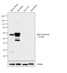
- Western blot analysis was performed on tissue extracts (30 µg lysate) of Mouse Brain (Lane 1), Rat Brain (Lane 2), Rat Liver (Lane 3) and Rat Kidney (Lane 4). The blot was probed with Anti- alpha Internexin Mouse Monoclonal Antibody (Product # 32-3600, 0.5 µg/mL) and detected by chemiluminescence using Goat anti-Mouse IgG (H+L) Superclonal™ Secondary Antibody, HRP conjugate (Product # A28177, 0.5 µg/mL, 1:4000 dilution). A 55 kDa band corresponding to alpha Internexin was observed in Mouse Brain, Rat Brain and not observed in other tissues which are documented to be alpha Internexin negative.
Supportive validation
- Submitted by
- Invitrogen Antibodies (provider)
- Main image

- Experimental details
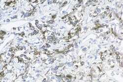
- Mouse anti-Alpha-Internexin (2E3) stained paraganglioma.
Supportive validation
- Submitted by
- Invitrogen Antibodies (provider)
- Main image

- Experimental details
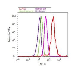
- Flow cytometry analysis of alpha-Internexin was performed using Neuro-2a cells. Cells were fixed with 70% ethanol for 10 minutes, permeabilized with 0.25% Triton™ X-100 for 20 minutes, and blocked with 5% BSA for 30 minutes at room temperature. Cells were labeled with alpha-Internexin Mouse Monoclonal Antibody (32-3600, red histogram) or with mouse isotype control (pink histogram) at 3-5 ug/million cells in 2.5% BSA. After incubation at room temperature for 2 hours, the cells were labeled with Alexa Fluor® 488 Rabbit Anti-Mouse Secondary Antibody (A11059) at a dilution of 1:400 for 30 minutes at room temperature. The representative 10,000 cells were acquired and analyzed for each sample using an Attune® Acoustic Focusing Cytometer. The purple histogram represents unstained control cells and the green histogram represents no-primary-antibody control..
Supportive validation
- Submitted by
- Invitrogen Antibodies (provider)
- Main image

- Experimental details
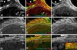
- NULL
 Explore
 Validate
 Learn
Learn Western blot
 ELISA
ELISA