Explore
 Validate
 Learn
Learn Western blot
 Immunocytochemistry
Antibody data
- Antibody Data
- Antigen structure
- References [0]
- Comments [0]
- Validations
- Western blot [4]
- Immunohistochemistry [2]
Submit
Validation data
Reference
Comment
Report error
- Product number
- NB300-140-0.1ml - Provider product page

- Provider
- Novus Biologicals
- Product name
- Mouse Monoclonal alpha-Internexin Antibody
- Antibody type
- Monoclonal
- Description
- Affinity purified.
- Reactivity
- Human, Mouse, Rat, Bovine, Porcine
- Host
- Mouse
- Antigen sequence
The C-terminal non-helical extensio
n of the protein.- Isotype
- IgG
- Vial size
- 0.1 ml
- Concentration
- 1 mg/ml
- Storage
- Store at 4C short term. Aliquot and store at -20C long term. Avoid freeze-thaw cycles.
No comments: Submit comment
Supportive validation
- Submitted by
- Novus Biologicals (provider)
- Main image

- Experimental details
- Simple Western: alpha-Internexin Antibody (2E3) [NB300-140] - Simple Western lane view shows a specific band for alpha Internexin in 0.5 mg/ml of SH-SY5Y lysate. This experiment was performed under reducing conditions using the 12-230 kDa separation system.

- Submitted by
- Novus Biologicals (provider)
- Main image
- Experimental details
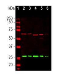
- Western Blot: alpha-Internexin Antibody (2E3) [NB300-140] - Analysis of different tissue lysates, dilution 1:10,000 in red : [1] protein standard (red), [2] rat brain, [3] rat spinal cord, [4] mouse brain, [5] mouse spinal cord, and [6] cow spinal cord lysate. Reveals the alpha-Internexin protein with apparent molecular weight of 64 - 66kDa. The same blot was simultaneously probed with chicken pAb to Calretinin, CPCA-Calretinin, dilution 1:1,000 in gree. A band at 29kDa corresponds to calretinin protein.

- Submitted by
- Novus Biologicals (provider)
- Main image

- Experimental details
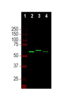
- Western Blot: alpha-Internexin Antibody (2E3) [NB300-140] - Analysis of spinal cord lysates from different species, dilution 1:5,000 in red : [1] protein standard (red), [2] mouse, [3] rat, and [4] cow spinal cord lysate. Antibody reveals the alpha-internexin protein with apparent molecular weight of 64 - 66kDa.
- Submitted by
- Novus Biologicals (provider)
- Main image

- Experimental details
- Western Blot: alpha-Internexin Antibody (2E3) [NB300-140] - Analysis of different tissue lysates using alpha-Internexin, dilution 1:10000 (Red): [1] protein standard, [2] rat brain, [3] rat spinal cord, [4] mouse brain, [5] mouse spinal cord, and [6] cow spinal cord lysate. The antibody reveals the alpha-Internexin protein with an apparent molecular weight of 64-66kDa, with some variability across species.

Supportive validation
- Submitted by
- Novus Biologicals (provider)
- Main image

- Experimental details
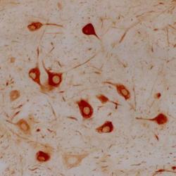
- Immunohistochemistry: alpha-Internexin Antibody (2E3) [NB300-140] - Rat facial nucleus before (left) and after (right) facial nerve lesion. Note the profound upregulation of a-internexin in the facial nucleus motor neurons.

- Submitted by
- Novus Biologicals (provider)
- Main image
- Experimental details
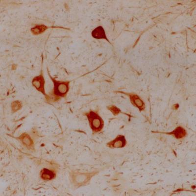
- Immunohistochemistry: alpha-Internexin Antibody (2E3) [NB300-140] - Section of rat facial nucleus 7 days following axotomy. These neurons are capable of regenerating their axons and also, concomitant with regeneration, strongly upregulate alpha-Internexin in their perikarya. Other central neurons which are not able to regenerate their axons do not upregulate this protein after axotomy and untreated facial neurons normally show only very low levels of alpha-Internexin. Both findings suggest that alpha-Internexin has a role in axonal regeneration.